Explore Your Local Site

Looks like you've landed on our   site. Let's take you home:    

Please note that the content and products on the    site might not be available in your region.


Choose the language:

  Homepage
Continue on the current website:  


Die aktuelle und künftige Rolle der künstlichen Intelligenz bei der Diagnose und Früherkennung von Brustkrebs

Brustkrebs

Brustkrebs ist weltweit die am häufigsten auftretende Krebsart bei Frauen (Sung et. al., 2021). Bei Frauen ist Brustkrebs für 1 von 4 Krebsfällen und 1 von 6 Krebstodesfällen verantwortlich, wobei er in der überwiegenden Mehrheit der Länder (159 von 185 Ländern) an erster Stelle der Inzidenz und in 110 Ländern an erster Stelle der Todesfälle steht (Sung et. al., 2021). Die meisten Fälle treten bei Frauen über 50 Jahren auf, es können jedoch auch jüngere Frauen betroffen sein. Weitere Risikofaktoren sind eine genetische Veranlagung, familiäre Vorbelastung, frühes Einsetzen der Menstruation, Hormonersatztherapie, Alkoholkonsum und Fettleibigkeit (Łukasiewicz et al., 2021).

Die Brust besteht aus milchproduzierenden Drüsenläppchen, einem System von Milchgängen und Fettgewebe (Bazira et al., 2021). Alle Brustkrebsarten haben ihren Ursprung in den Zellen, die die terminalen duktulo-lobulären Einheiten (die funktionelle Einheit der Brust) der Sammelgänge auskleiden. Die häufigste Form des männlichen Brustkrebses ist das invasive duktale Karzinom, das in den Milchgängen beginnt und in das umliegende Gewebe eindringt (Harbeck et al., 2019). An der Entstehung von Brustkrebs sind genetische Mutationen beteiligt, die eine unkontrollierte Zellvermehrung verursachen, sowie die Gene BRCA1 und BRCA2, die an der DNA-Reparatur beteiligt sind (Harbeck et al., 2019). Östrogen- und Progesteronrezeptoren spielen eine wichtige Rolle in der Pathophysiologie; alle Patienten mit Tumoren, die diese Rezeptoren exprimieren, sollten eine Hormontherapie erhalten, um die Östrogenrezeptoraktivität zu blockieren (Harbeck et al., 2019).

Brustkrebs kann auf verschiedene Weise in Erscheinung treten. Das häufigste klinische Merkmal ist ein Knoten in der Brust, Veränderungen in der Größe der Brustwarze, Ausfluss aus der Brustwarze und Hautveränderungen sowie eine Infektion und/oder Entzündung der Brust (Koo et al., 2017). Brustkrebs im Frühstadium ist häufig asymptomatisch, was die Bedeutung von Routineuntersuchungen unterstreicht (Kalager et al., 2010).

Brustkrebs wird in der Regel durch ein Screening oder ein Symptom (Schmerzen oder tastbarer Knoten) diagnostiziert, das eine diagnostische Untersuchung erfordert (McDonald et al., 2016). Dazu kommen bildgebende Verfahren, um nach Anomalien zu suchen und sie genauer zu charakterisieren (McDonald et al., 2016). Eine Brustbiopsie wird in der Regel durchgeführt, um das Vorhandensein von Krebs zu bestätigen, wenn der Verdacht besteht, und um den spezifischen Typ der Läsion zu bestimmen, wenn es sich um Krebs handelt (McDonald et al., 2016). Brustkrebs wird anhand der Ausdehnung des Tumors, der Ausbreitung auf nahe gelegene Lymphknoten, der Ausbreitung auf entfernte Stellen, des Östrogenrezeptorstatus, des Progesteronrezeptorstatus, des HER2-Status und des Grades der Krebserkrankung eingestuft (McDonald et al., 2016).

Es gibt verschiedene Arten von Brustkrebs, und die Behandlung kann je nach den molekularen Merkmalen der Krankheit einer Patientin, dem Stadium, der Krebsart und dem Rezeptorstatus variieren (Hong & Xu, 2022). Die Behandlung umfasst in der Regel eine Kombination verschiedener Methoden und ein multidisziplinäres Team von Gesundheitsexperten (Hong & Xu, 2022). Die chirurgischen Optionen reichen von brusterhaltenden Verfahren bis hin zur Mastektomie, bei der die gesamte Brust entfernt wird (Hong & Xu, 2022). Die Entfernung von Lymphknoten kann auch notwendig sein, um das Ausmaß der Krebsausbreitung zu beurteilen (Hong & Xu, 2022). Die Strahlentherapie wird häufig nach brusterhaltender Therapie oder Mastektomie (mit Risikofaktoren) eingesetzt (Hong & Xu, 2022). Die systemische Chemotherapie kann je nach Situation vor oder nach der Operation verabreicht werden (Hong & Xu, 2022). Hormonrezeptor-positiver Brustkrebs kann mit Medikamenten behandelt werden, die die Wirkung von Östrogen und Progesteron blockieren. Die Immuntherapie ist eine neue Behandlungsoption für bestimmte Brustkrebsarten, die dem Immunsystem hilft, Krebszellen zu erkennen und anzugreifen (Hong & Xu, 2022).

Bildgebungstechniken

Digitale Mammografie

Die digitale Mammografie ist die am häufigsten verwendete Technik für das Brustkrebs-Screening. Es handelt sich um eine zweidimensionale Summationstechnik, bei der die von einer Röntgenröhre ausgesandten Röntgenstrahlen in unterschiedlichem Maße von Geweben absorbiert und von einem Detektor auf der anderen Seite gemessen werden. Dichteres Gewebe erscheint auf den resultierenden Bildern heller als weniger dichtes Gewebe. Die Brüste werden bei der Bildaufnahme zusammengedrückt, um das Brustgewebe auf eine größere Fläche zu verteilen (Ikeda, 2011a). Dadurch wird die Überlappung zwischen verschiedenen Komponenten des Brustgewebes reduziert, die Streuung der durchgehenden Röntgenstrahlen verringert und der Kontrast verbessert. In der Regel werden zwei Ansichten von jeder Brust aufgenommen – kraniokaudal (CC) und mediolateral (MLO) (Ikeda, 2011a).

mammography

Die digitale Mammografie ist eine schnelle und nützliche Technik für das Brustkrebsscreening, aber sie hat auch ihre Nachteile (Ikeda, 2011a). Die Kompression der Brust kann schmerzhaft sein, und die Überlappung verschiedener Gewebe trotz der Kompression führt häufig zu Artefakten (Ikeda, 2011a). Der obere innere Quadrant der Brust, der weniger beweglich ist, da er fest mit der Brustwand verbunden ist, ist in der Mammografie besonders schwer darzustellen (Ikeda, 2011a). Auch in Brüsten mit einem hohen Anteil an dichtem Gewebe kann Krebs in der Mammografie sehr schwer zu erkennen sein (Ikeda, 2011a).

Digitale Brusttomosynthese

Bei der digitalen Brusttomosynthese (DBT) werden die Bilder mithilfe einer Röntgenröhre aufgenommen, die eine Bogenbewegung ausführt. Dünne Schichten werden rekonstruiert und ermöglichen eine 3D-Bildgebung, die den Einfluss von überlappendem Brustgewebe minimieren soll und besonders nützlich für die Darstellung von Brustläsionen ist, die sich in einem heterogenen und dichten Brustparenchym befinden. Eine Studie ergab, dass die DBT bei der Erkennung von Brustkrebs empfindlicher ist als die digitale Mammografie (DM). Die DBT kann mit der DM kombiniert werden, und eine Studie ergab, dass die Kombination dieser Techniken die Erkennung von Brustkrebs verbessert (Alabousi et al., 2020; Lei et al., 2014; Skaane et al., 2019), und die Kombination mit der Mammografie möglich ist. Die DBT dauert jedoch länger als die Mammografie und ist mit Bewegungs- und anderen Artefakten behaftet (Tirada et al., 2019).

Ultraschall

Beim diagnostischen Ultraschall sendet ein Schallkopf Hochfrequenz-Schallwellen aus, die sich durch das Gewebe bewegen, an diesem abprallen und „Echos“ erzeugen, die auf den Schallkopf zurückgeworfen und von diesem erkannt werden. Diese Echoaufnahmen werden dann verarbeitet, um Echtzeitbilder auf einem Monitor zu erstellen, die auf der Zeit basieren, die die Ultraschallwellen für den Weg zum Gewebe und zurück benötigen. Es handelt sich um eine sichere und relativ kostengünstige Technik, die häufig als Ergänzung zur Mammografie eingesetzt wird (Ikeda, 2011b), insbesondere zur weiteren Beurteilung eines tastbaren oder mammografischen Befunds.

ultrasound

Sie kann sogar als primäre Screening-Methode bei Frauen unter 30 Jahren oder bei schwangeren oder stillenden Frauen eingesetzt werden (Dixon, 2008; Ikeda, 2011b). Ultraschall ist sehr nützlich, um zu klären, ob eine Masse zystisch oder solide ist, welche Art von Rändern sie hat und ob sie vaskulär ist (Dixon, 2008; Ikeda, 2011b). Sie hilft auch, andere Massen und verdächtige axilläre Lymphknoten zu entdecken (Dixon, 2008; Ikeda, 2011b). Der größte Nachteil ist, dass die Qualität der Untersuchung stark bedienerabhängig ist (Dixon, 2008; Ikeda, 2011b).

Magnetresonanztomographie

Mithilfe eines starken Magnetfelds und einer Reihe von Hochfrequenzwellen richtet die Magnetresonanztomographie (MRT) die Wasserstoffkerne im Gewebe gleichmäßig aus, um detaillierte Querschnittsbilder des Körpers zu erstellen (Daniel & Ikeda, 2011; Mann et al., 2019). Da Gewebe mit unterschiedlicher Zusammensetzung auf diese Störung unterschiedlich reagieren, kann die MRT selbst kleinste Unterschiede zwischen verschiedenen Weichgewebetypen sehr gut nachweisen und gilt als die genaueste Methode zur Diagnose von Brustkrebs (Daniel & Ikeda, 2011; Mann et al., 2019). Sie wird hauptsächlich für das Screening von Hochrisikopatienten auf der Grundlage genetischer oder erworbener Risikofaktoren verwendet (Daniel & Ikeda, 2011).

MRI

Für die MRT der Brust sind spezielle Brustspulen erforderlich, die die Hochfrequenzwellen aussenden und das erzeugte Signal empfangen. Die Bilder werden häufig mit einer räumlichen Auflösung von 1 mm in der Ebene, einer Schichtdicke von weniger als 3 mm und einer Unterdrückung des Fettgewebesignals aufgenommen. Zu den häufig verwendeten Sequenzen gehören T2-gewichtete Bilder, diffusionsgewichtete Bilder und dynamische kontrastverstärkte MRT. Um falsch-positive Ergebnisse aufgrund unspezifischer Veränderungen des Brustparenchyms zu vermeiden, wird die Untersuchung am besten zwischen Tag 7 und 13 des Menstruationszyklus durchgeführt (Daniel & Ikeda, 2011). Anders als bei der Mammografie werden bei der MRT keine ionisierenden Strahlen eingesetzt und dreidimensionale Bilder erzeugt, die die Erkennung sehr kleiner Läsionen ermöglichen (DeMartini & Lehman, 2008; Shahid et al., 2016). Die MRT ermöglicht auch eine detailliertere Beurteilung der Brustwand als die Mammografie und der Ultraschall (DeMartini & Lehman, 2008). Zu den Nachteilen der Brust-MRT gehören die geringe Erkennungsfähigkeit von Mikroverkalkungen, die hohen Kosten und die Tatsache, dass sie bei Personen mit bestimmten Metallimplantaten kontraindiziert ist (Daniel & Ikeda, 2011).

Screening- und Diagnoseherausforderungen

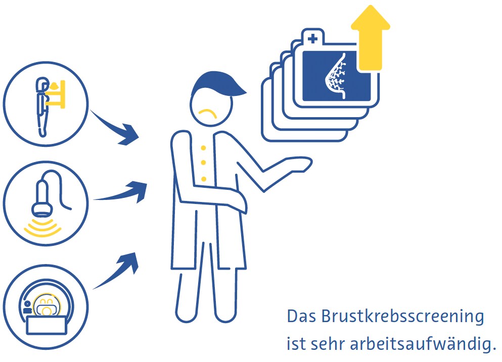
Trotz der Belege für den allgemeinen Nutzen des Brustkrebsscreenings (Dibden et al., 2020; Kalager et al., 2010; Tabár et al., 2019) ist es mit mehreren technischen und logistischen Herausforderungen behaftet. Bei mehr als der Hälfte der Frauen, die 10 Jahre lang jährlich untersucht werden, fällt der Test falsch positiv aus (Hubbard et al., 2011). Dies hat weitreichende und signifikante Folgen, einschließlich der physischen und emotionalen Belastung durch unnötige Biopsien und erhöhte Gesundheitskosten (Nelson, Pappas, et al., 2016; Ong & Mandl, 2015). Auch beim Screening wird Brustkrebs häufig übersehen, insbesondere bei Frauen mit dichter Brust (Banks et al., 2006).
breast cancer

Das Brustkrebsscreening erfordert hochqualifizierte Fachkräfte, darunter Radiologen und Röntgenassistenten, an denen es derzeit weltweit mangelt (Moran & Warren-Forward, 2012; Rimmer, 2017; Wing & Langelier, 2009). Dieses Problem wird durch die Tatsache verschärft, dass in vielen europäischen Ländern der Standard der Versorgung beim Brustscreening darin besteht, dass jede Auswertung von zwei Radiologen unabhängig voneinander durchgeführt wird (Giordano et al., 2012), und dass in bestimmten Ländern, wie z. B. in den USA, die Hürden für die Qualifikation zur Interpretation von Mammogrammen aufgrund strenger beruflicher Zulassungsstandards hoch sind (Food and Drug Administration, 2001).

Außerdem gibt es weltweit erhebliche Hindernisse für die Inanspruchnahme des Brustkrebsscreenings. Dazu gehören der fehlende oder schwierige Zugang zu Früherkennungsprogrammen, mangelndes Wissen oder Missverständnisse über den Nutzen dieser Programme sowie soziale und kulturelle Barrieren (Mascara & Constantinou, 2021).

Rolle der künstlichen Intelligenz

Technische Verbesserungen

Bisher gibt es nur wenige veröffentlichte Studien, die den Einsatz von KI zur technischen Verbesserung von Brustuntersuchungen direkt untersucht haben. Eine kommerziell erhältliche Anwendung gibt dem Röntgenpersonal in Echtzeit Rückmeldung über die korrekte Positionierung der Patientin bei Mammografien. (Volpara Gesundheit, 2022). Andere KI-Anwendungen konzentrieren sich auf die Verringerung der Strahlendosis (J. Liu et al., 2018), die Verbesserung der Bildrekonstruktion (Kim et al., 2016) und die Reduzierung von Rauschen und Artefakten bei der DBT (Garrett et al., 2018).

Beim Brustkrebsscreening wird die DBT häufig mit der digitalen Mammografie kombiniert, wodurch sich die Strahlendosis für die Patientinnen und Patienten verdoppelt (Svahn et al., 2015). Um dies zu vermeiden, hat das Interesse an der Erstellung synthetischer Mammogramme aus DBT-Daten zugenommen (Chikarmane et al., 2023). In einer großen prospektiven norwegischen Studie war die Genauigkeit der DBT in Kombination mit digitaler Mammografie oder synthetischer Mammografie bei der Brustkrebserkennung sehr ähnlich (Skaane et al., 2019). Jüngste Studien haben die Verbesserung der Qualität der synthetischen Mammografie durch KI untersucht und vielversprechende Ergebnisse erzielt (Balleyguier et al., 2017; James et al., 2018).

Diagnostische Verbesserungen

Beurteilung der Brustdichte

Dichtes Brustgewebe, das auf der Mammografie sichtbar ist, stellt fibroglanduläres Gewebe dar. Frauen mit dichten Brüsten haben ein 2- bis 4-fach höheres Brustkrebsrisiko als Frauen mit Brüsten mit mehr fetthaltigem Brustgewebe (Byrne et al., 1995; Duffy et al., 2018; Torres-Mejía et al., 2005). Darüber hinaus ist die Sensitivität der Mammografie für Brustkrebs in dichten Brüsten um 20-30 % geringer als in weniger dichten Brüsten (Lynge et al., 2019). Der Standard für die Beurteilung der Brustdichte ist die BI-RADS-Klassifikation (Berg et al., 2000).

Mehrere große Studien haben das Potenzial für die automatische Bewertung der Brustdichte auf Mammo-grammen mithilfe von KI-basierten Tools untersucht. Ein konvolutionales neuronales Netz (aus dem Englischen für CNN: Convolutional Neural Network) das auf 14.000 Mammogrammen trainiert und auf fast 2000 Mammo-grammen getestet wurde, klassifizierte die Brustdichte entweder als „verstreut dicht“ oder „heterogen dicht“ mit einer Fläche unter der Kurve (AUC) von 0,93 (Mo-hamed et al., 2018). In einer anderen Studie wurde ein CNN verwendet, das sowohl eine binäre als auch eine vierfache BI-RADSKlassifizierung durchführen kann und mit mehr als 40 000 Mammogrammen trainiert wurde (Lehman et al., 2019). In einem Testdatensatz von mehr als 8.000 Mammogrammen fanden sie einen hohen Grad an Übereinstimmung bei der Brustdichte zwischen dem Algorithmus und einzelnen Radiologen (kappa = 0,67) sowie dem Konsens von fünf Radiologen (kappa = 0,78) (Lehman et al., 2019).

Brustkrebserkennung

In einer systematischen Übersichtsarbeit, die 82 Studien mit KI zur Brustkrebserkennung mit verschiedenen Referenzstandards umfasste, fanden die Autoren eine AUC von 0,87 für Studien mit Mammografie, 0,91 mit Ultraschall, 0,91 mit DBT und 0,87 mit MRT (Aggarwal et al., 2021). Dies sind vielversprechende Ergebnisse, doch ein direkter Vergleich zwischen KI-basierten Algorithmen und Radiologen zeigt, dass es noch Raum für Verbesserungen gibt. In einer anderen systematischen Überprüfung von Studien, in denen entweder die Histopathologie oder die Nachbeobachtung (bei Screening-negativen Frauen) als Referenz herangezogen wurde, waren 94 % der 36 identifizierten CNNs weniger genau als ein einzelner Radiologe, und alle waren weniger genau als der Konsens von zwei oder mehr Radiologen, wenn sie als eigenständiges System verwendet wurden (Freeman et al., 2021). Die derzeitigen Erkenntnisse sprechen daher nicht für den Einsatz von KI als alleinige Strategie zur Brustkrebserkennung.

Brustkrebsvorhersage

KI hat sich als vielversprechend für die Vorhersage des Brustkrebsrisikos auf der Grundlage von Screening- Mammogrammen erwiesen, entweder durch eine bessere Bewertung der Brustdichte, einem etablierten Risikofaktor für Brustkrebs (Duffy et al., 2018), oder durch die Erkennung feinster Bildgebungsmerkmale, die Vorboten von Krebs sind (Batchu et al., 2021). In mehreren Studien wurden KI-basierte Modelle verwendet, um das Risiko, in Zukunft an Brustkrebs zu erkranken, auf der Grundlage von Mammogrammen vorherzusagen (Batchu et al., 2021; Geras et al., 2019).

Ein CNN, das auf fast 1 000 000 Mammografie-Bildern trainiert wurde, zeigte eine AUC von 0,65 für die Vorhersage der zukünftigen Entwicklung von Brustkrebs im Vergleich zu 0,57-0,60 für konventionelle Mammografie- basierte Brustdichte-Scores (Dembrower, Liu, et al., 2020). Eine kleinere Studie ergab einen AUC-Wert von 0,73 für eine CNN-basierte Methode zur Vorhersage von Brustkrebs aus normalen Mammografiebildern. (Arefan et al., 2020). Ein anderer Deep-Learning-Algorithmus zeigte eine AUC von 0,82 für die Vorhersage von Intervallkrebs (Krebs, der innerhalb von 12 Monaten nach einer negativen Mammografie entdeckt wurde) im Vergleich zu 0,65 für die visuelle BI-RADS-Bewertung der Brustdichte (Hinton et al., 2019). Ein anderes Deep- Learning-basiertes Modell, das sowohl Risikofaktoren als auch mammografische Befunde zur Vorhersage des Brustkrebsrisikos einbezog, hatte eine AUC von bis zu 0,7 und übertraf damit die Genauigkeit von Vorhersagemodellen, die nur auf Risikofaktoren oder mammografischen Befunden basieren. (Yala, Lehman, et al., 2019).

Effizienzsteigerungen

Die schiere Menge an mammografischen Untersuchungen und der Mangel an geschulten Radiologen haben Effizienzsteigerungen zu einem der interessantesten Forschungsbereiche für den Einsatz von KI bei Brustkrebs gemacht.

In einer Studie simulierten die Autoren einen Arbeitsablauf, bei dem Mammogramme von einem Radiologen und einem Deep-Learning-Modell interpretiert wurden, wobei die Entscheidung als endgültig galt, wenn beide übereinstimmten (McKinney et al., 2020). Ein zweiter Radiologe wurde nur bei Nichtübereinstimmung hinzugezogen, was mit einer 88%igen Verringerung des Arbeitsaufwands für den zweiten Radiologen und einem negativen Vorhersagewert von über 99,9 % verbunden war (McKinney et al., 2020).

In einer erstmalig durchgeführten großen randomisierten klinischen Studie in Schweden wurden etwa 80 000 Frauen ausgewählt, deren Screening-Mammogramme entweder einer Vorauswertung durch einen CNN unterzogen wurden oder nicht (Lång et al., 2023). In der Untersuchungsgruppe wurden nur Mammogramme, denen eine hohe Malignitätswahrscheinlichkeit zugewiesen wurde, doppelt ausgewertet (der Rest wurde von einem Radiologen ausgewertet), und die Ergebnisse wurden mit der herkömmlichen doppelten Auswertung ohne Hilfe des Algorithmus verglichen. In einer Zwischenanalyse der Daten der 80.000 Frauen zeigten beide Studienarme eine identische Falsch-Positiv-Rate von 1,5 %. Der positive Vorhersagewert der Befundung betrug in der Untersuchungsgruppe 28,3 % und in der Kontrollgruppe 24,8 %, und der Arbeitsaufwand wurde durch die Strategie um 44,3 % reduziert (Lång et al., 2023).

In anderen Studien wurde die KI zur Voruntersuchung von Mammogrammen eingesetzt, wobei diejenigen mit einer geringen Krebswahrscheinlichkeit aussortiert wurden und nur diejenigen mit einer hohen Krebswahrscheinlichkeit einem Radiologen vorgelegt wurden. In einer Studie aus den USA wurde zu diesem Zweck ein simulierter Arbeitsablauf verwendet, bei dem ein CNN auf mehr als 212 000 Mammogrammen trainiert und auf über 26 000 getestet wurde (Yala, Schuster, et al., 2019). Der Arbeitsablauf unter Verwendung des Algorithmus hatte eine nicht unterlegene Sensitivität für Brustkrebs (90,1 % vs. 90,6 %) und eine etwas höhere Spezifität im Vergleich zu allein arbeitenden Radiologen (94,2 % gegenüber 93,5 %) und war mit einem um 19,3 % geringeren Arbeitsaufwand verbunden (Yala, Schuster, et al., 2019). Eine kleinere Studie aus Spanien ergab eine Verringerung des Arbeitsaufwands um 72,5 %, wenn die KI nur DBT-Fälle mit hohem Risiko für die Auswertung durch einen zweiten Radiologen auswählte, und um 29,7 %, wenn die KI nur DBT-Studien mit hohem Risiko für die Auswertung durch einen zweiten Radiologen auswählte, im Vergleich zu herkömmlichen Mammografie-Workflows mit doppelter Auswertung (Raya-Povedano et al., 2021). Sie stellten außerdem fest, dass diese Strategie, bei der KI zur Auswahl von Mammografie- und DBT-Fällen mit hohem Risiko für eine zweite Auswertung eingesetzt wird, im Vergleich zu den standardmäßigen Mammografie- und DBT- Workflows mit doppelter Auswertung eine nicht schlechtere Sensitivität aufweist (Raya-Povedano et al., 2021). In einer schwedischen Studie ergab eine ähnliche Strategie unter Verwendung eines kommerziell verfügbaren KI-Algorithmus eine Falsch-Negativ-Rate von nicht mehr als 4 %, und der Algorithmus zeigte die Fähigkeit, möglicherweise 71 zusätzliche Krebsfälle pro 1000 Untersuchungen zu entdecken, mehr als eine doppelte Bewertung auf negative Befunde durch menschliche Radiologen bei Patienten, die der KI-Algorithmus als sehr risikoreich einstufte (Dembrower, Wåhlin, et al., 2020).

In einer Studie mit über einer Million Mammogrammen an acht Screening-Standorten und von drei Geräteherstellern hat ein kommerziell erhältlicher Deep-Learning- Algorithmus auf der Grundlage von Bewertungen mit hoher Konfidenz 63 % der Untersuchungen so eingestuft, dass sie nicht weiter verfolgt werden sollten (Leibig et al., 2022). Der Rest der Untersuchungen, bei denen der Algorithmus eine geringe Konfidenz aufwies, wurde den Radiologen vorgelegt. Diese Strategie verbesserte die Sensitivität der Radiologen (im Vergleich zur nicht unterstützten Auswertung) um 2,6-4 % und die Spezifität um 0,5-1,0 % (Leibig et al., 2022).
 

Herausforderungen und zukünftige Wege

Mehrere ethische, technische und methodische Herausforderungen, die mit dem Einsatz von KI in der Brustkrebsvorsorge verbunden sind, bilden einen Rahmen für die künftige Forschung zu diesem Thema (Hickman et al., 2021).

Die meisten KI-basierten Tools haben sich bisher auf die digitale Mammografie konzentriert (Aggarwal et al., 2021), aber andere Untersuchungstechniken wie DBT und MRT haben einzigartige Vorteile (Alsheik et al., 2019; Mann et al., 2019) und werden in Zukunft wahrscheinlich eine größere Rolle beim Brustkrebs-Screening spielen. Da es sich bei DBT und MRT jedoch um tomografische Verfahren handelt, die dreidimensionale Ergebnisse liefern, erfordert die Verarbeitung mit KI-basierten Tools mehr Speicherplatz und Rechenleistung (Prevedello et al., 2019).

Inzidenz, Verlauf und Ergebnis von Brustkrebs hängen mit verschiedenen soziodemografischen Faktoren zusammen, darunter ethnische Herkunft und Zugehörigkeit (Hirko et al., 2022; Hu et al., 2019; Martini et al., 2022). Das Training von KI-basierten Hilfsmitteln mit Datensätzen, die ein breites Spektrum der Menschen repräsentieren, ist der Schlüssel, um sicherzustellen, dass sie verallgemeinert werden können und so vielen Menschen wie möglich nützen.

Die allgemeine Leistung der KI bei der Erkennung von Brustkrebs ist beeindruckend. Es ist jedoch hervorzuheben, dass in einer Studie nicht nachgewiesen werden konnte, dass die Sensitivität der KI bei der Erkennung von Brustkrebs derjenigen von Radiologen nicht unterlegen ist (Lauritzen et al., 2022). Darüber hinaus ist die Qualität der Nachweise in vielen Studien zu diesem Thema fraglich. Eine systematische Überprüfung der Genauigkeit von KI-basierten Tools für die Brustkrebserkennung ergab mehrere Bereiche mit Verbesserungspotenzial (Freeman et al., 2021). Bei der Überprüfung wurden keine prospektiven Studien gefunden, und die identifizierten Studien waren von schlechter methodischer Qualität. Insbesondere stellten die Autoren fest, dass kleinere Studien positivere Ergebnisse zeigten, die sich in größeren Studien nicht wiederholen ließen. In einer anderen systematischen Übersichtsarbeit wurde nur in etwa einem Zehntel der Studien ein externer Datensatz zur Validierung herangezogen, in keiner Studie wurde eine vorgegebene Stichprobengröße berechnet, und es wurden schwerwiegende Probleme mit Selektionsverzerrungen und unangemessenen Referenzstandards festgestellt (Aggarwal et al., 2021). Diese methodischen Probleme können in Zukunft möglicherweise durch die Einführung großer offener Datenbestände (Nguyen et al., 2023) und die verstärkte Einhaltung von Leitlinien für die Durchführung KI-gestützter medizinischer Forschung (Lekadir et al., 2021; X. Liu et al., 2020) entschärft werden.
 

Fazit

Die Integration von künstlicher Intelligenz in Mammographie-Screening Programme zur Brustkrebs- Früherkennung verspricht, die Bildqualität, Effi-zienz und Vorhersage des zukünftigen Brustkrebsrisikos zu verbessern. Bei der Erkennung von Brustkrebs bei Screening-Untersuchungen zeigt sich, dass künstliche Intelligenz am besten funktioniert, wenn sie gemeinsam mit Radiologen eingesetzt wird. Die laufende Forschung ist von entscheidender Bedeutung, um die mit dem Einsatz von KI in der Brustkrebsvorsorge verbundenen Herausforderungen zu bewältigen, einschließlich der Ausweitung ihrer Anwendungen über die Mammografie hinaus und der Gewährleistung ihres ethischen und ver-antwortungsvollen Einsatzes. Mit der kontinuierlichen Weiterentwicklung von KIAnwendungen birgt die Zukun-ft der Brustkrebsvorsorge ein immenses Potenzial für eine bessere Zugänglichkeit, ein frühzeitiges Intervention und letztlich bessere Ergebnisse für die Patientinnen.

AI

Literaturverzeichnis 

Aggarwal, R., Sounderajah, V., Martin, G., Ting, D. S. W., Karthikesalingam, A., King, D., Ashrafian, H., & Darzi, A. (2021). Diagnostic accuracy of deep learning in medical imaging: a systematic review and meta-analysis. NPJ Digital Medicine, 4(1), 65.

Alabousi, M., Zha, N., Salameh, J.-P., Samoilov, L., Sharifabadi, A. D., Pozdnyakov, A., Sadeghirad, B., Freitas, V., McInnes, M. D. F., & Alabousi, A. (2020). Digital breast tomosynthesis for breast cancer detection: a diagnostic test accuracy systematic review and meta-analysis. European Radiology, 30(4), 2058–2071.

Alkabban FM and Ferguson T. (2022). Breast Cancer, 2022. In: StatPearls [Internet]. Treasure Island (FL): StatPearls Publishing.

Alsheik, N. H., Dabbous, F., Pohlman, S. K., Troeger, K. M., Gliklich, R. E., Donadio, G. M., Su, Z., Menon, V., & Conant, E. F. (2019). Comparison of Resource Utilization and Clinical Outcomes Following Screening with Digital Breast Tomosynthesis Versus Digital Mammography: Findings From a Learning Health System. Academic Radiology, 26(5), 597–605.

Arefan, D., Mohamed, A. A., Berg, W. A., Zuley, M. L., Sumkin, J. H., & Wu, S. (2020). Deep learning modeling using normal mammograms for predicting breast cancer risk. Medical Physics, 47(1), 110–118.

Balleyguier, C., Arfi-Rouche, J., Levy, L., Toubiana, P. R., Cohen-Scali, F., Toledano, A. Y., & Boyer, B. (2017). Improving digital breast tomosynthesis reading time: A pilot multi-reader, multi-case study using concurrent Computer-Aided Detection (CAD). European Journal of Radiology, 97, 83–89.

Banks, E., Reeves, G., Beral, V., Bull, D., Crossley, B., Simmonds, M., Hilton, E., Bailey, S., Barrett, N., Briers, P., English, R., Jackson, A., Kutt, E., Lavelle, J., Rockall, L., Wallis, M. G., Wilson, M., & Patnick, J. (2006). Hormone replacement therapy and false positive recall in the Million Women Study: patterns of use, hormonal constituents and consistency of effect. Breast Cancer Research: BCR, 8(1), R8.

Batchu, S., Liu, F., Amireh, A., Waller, J., & Umair, M. (2021). A Review of Applications of Machine Learning in Mammography and Future Challenges. Oncology, 99(8), 483–490. https://doi.org/10.1159/000515698

Bazira, P. J., Ellis, H., & Mahadevan, V. (2022). Anatomy and physiology of the breast. Surgery, 40(2), 79–83. https://doi.org/10.1016/j.mpsur.2021.11.015

Berg, W. A., Campassi, C., Langenberg, P., & Sexton, M. J. (2000). Breast Imaging Reporting and Data System: interand intraobserver variability in feature analysis and final assessment. AJR. American Journal of Roentgenology, 174(6), 1769–1777.

Boyd, N. F., Guo, H., Martin, L. J., Sun, L., Stone, J., Fishell, E., Jong, R. A., Hislop, G., Chiarelli, A., Minkin, S., & Yaffe, M. J. (2007). Mammographic density and the risk and detection of breast cancer. The New England Journal of Medicine, 356(3), 227–236.

Bray, F., Ferlay, J., Soerjomataram, I., Siegel, R. L., Torre, L. A., & Jemal, A. (2018). Global cancer statistics 2018: GLOBOCAN estimates of incidence and mortality worldwide for 36 cancers in 185 countries. CA: A Cancer Journal for Clinicians, 68(6), 394–424.

Breast Cancer Statistics. (2020, October 27). Susan G. Komen®. https://www.komen.org/breast-cancer/facts-statistics/breastcancer- statistics/

Byrne, C., Schairer, C., Wolfe, J., Parekh, N., Salane, M., Brinton, L. A., Hoover, R., & Haile, R. (1995). Mammographic features and breast cancer risk: effects with time, age, and menopause status. Journal of the National Cancer Institute, 87(21), 1622–1629.

Chikarmane, S. A., Offit, L. R., & Giess, C. S. (2023). Synthetic Mammography: Benefits, Drawbacks, and Pitfalls. Radiographics: A Review Publication of the Radiological Society of North America, Inc, 43(10), e230018. https://doi.org/10.1148/rg.230018

Daniel, B. L., & Ikeda, D. M. (2011). Chapter 7 - Magnetic Resonance Imaging of Breast Cancer and MRI-Guided Breast Biopsy. In D. M. Ikeda (Ed.), Breast Imaging (Second Edition) (pp. 239–296). Mosby.

DeMartini W & Lehman C, (2008). Top Magn Reson Imaging, Jun;19(3):143-50.

Dembrower, K., Liu, Y., Azizpour, H., Eklund, M., Smith, K., Lindholm, P., & Strand, F. (2020). Comparison of a Deep Learning Risk Score and Standard Mammographic Density Score for Breast Cancer Risk Prediction. Radiology, 294(2), 265–272.

Dembrower, K., Wåhlin, E., Liu, Y., Salim, M., Smith, K., Lindholm, P., Eklund, M., & Strand, F. (2020). Effect of artificial intelligence-based triaging of breast cancer screening mammograms on cancer detection and radiologist workload: a retrospective simulation study. The Lancet. Digital Health, 2(9), e468–e474.

Dibden, A., Offman, J., Duffy, S. W., & Gabe, R. (2020). Worldwide Review and Meta-Analysis of Cohort Studies Measuring the Effect of Mammography Screening Programmes on Incidence-Based Breast Cancer Mortality. Cancers, 12(4). https://doi.org/10.3390/cancers12040976

Duffy, S. W., Morrish, O. W. E., Allgood, P. C., Black, R., Gillan, M. G. C., Willsher, P., Cooke, J., Duncan, K. A., Michell, M. J., Dobson, H. M., Maroni, R., Lim, Y. Y., Purushothaman, H. N., Suaris, T., Astley, S. M., Young, K. C., Tucker, L., & Gilbert, F. J. (2018). Mammographic density and breast cancer risk in breast screening assessment cases and women with a family history of breast cancer. European Journal of Cancer, 88, 48–56.

Food and Drug Administration. (2001). The Mammography Quality Standards Act Final Regulations: Preparing for MQSA Inspections; Final Guidance for Industry and FDA.

Freeman, K., Geppert, J., Stinton, C., Todkill, D., Johnson, S., Clarke, A., & Taylor-Phillips, S. (2021). Use of artificial intelligence for image analysis in breast cancer screening programmes: systematic review of test accuracy. BMJ, 374, n1872.

Freer, P. E. (2015). Mammographic breast density: impact on breast cancer risk and implications for screening. Radiographics: A Review Publication of the Radiological Society of North America, Inc, 35(2), 302–315.

Garrett, J. W., Li, Y., Li, K., & Chen, G.-H. (2018). Reduced anatomical clutter in digital breast tomosynthesis with statistical iterative reconstruction. Medical Physics, 45(5), 2009–2022.

Geras, K. J., Mann, R. M., & Moy, L. (2019). Artificial Intelligence for Mammography and Digital Breast Tomosynthesis: Current Concepts and Future Perspectives. Radiology, 293(2), 246–259. https://doi.org/10.1148/radiol.2019182627

Giordano, L., von Karsa, L., Tomatis, M., Majek, O., de Wolf, C., Lancucki, L., Hofvind, S., Nyström, L., Segnan, N., Ponti, A., Eunice Working Group, Van Hal, G., Martens, P., Májek, O., Danes, J., von Euler-Chelpin, M., Aasmaa, A., Anttila, A., Becker, N., … Suonio, E. (2012). Mammographic screening programmes in Europe: organization, coverage and participation. Journal of Medical Screening, 19 Suppl 1, 72–82.

Harbeck, N., Penault-Llorca, F., Cortes, J., Gnant, M., Houssami, N., Poortmans, P., Ruddy, K., Tsang, J., & Cardoso, F. (2019). Breast cancer. Nature Reviews. Disease Primers, 5(1), 66. https://doi.org/10.1038/s41572-019-0111-2

Hickman, S. E., Baxter, G. C., & Gilbert, F. J. (2021). Adoption of artificial intelligence in breast imaging: evaluation, ethical constraints and limitations. British Journal of Cancer, 125(1), 15–22.

Hinton, B., Ma, L., Mahmoudzadeh, A. P., Malkov, S., Fan, B., Greenwood, H., Joe, B., Lee, V., Kerlikowske, K., & Shepherd, J. (2019). Deep learning networks find unique mammographic differences in previous negative mammograms between interval and screen-detected cancers: a case-case study. Cancer Imaging: The Official Publication of the International Cancer Imaging Society, 19(1), 41.

Hirko, K. A., Rocque, G., Reasor, E., Taye, A., Daly, A., Cutress, R. I., Copson, E. R., Lee, D.-W., Lee, K.-H., Im, S.-A., & Park, Y. H. (2022). The impact of race and ethnicity in breast cancer-disparities and implications for precision oncology.BMC Medicine, 20(1), 72.

Hong, R., & Xu, B. (2022). Breast cancer: an up-to-date review and future perspectives. Cancer Communications, 42(10), 913–936. https://doi.org/10.1002/cac2.12358

Hubbard, R. A., Kerlikowske, K., Flowers, C. I., Yankaskas, B. C., Zhu, W., & Miglioretti, D. L. (2011). Cumulative probability of false-positive recall or biopsy recommendation after 10 years of screening mammography: a cohort study.Annals of Internal Medicine, 155(8), 481–492.

Hu, K., Ding, P., Wu, Y., Tian, W., Pan, T., & Zhang, S. (2019). Global patterns and trends in the breast cancer incidence and mortality according to sociodemographic indices: an observational study based on the global burden of diseases. BMJ Open, 9(10), e028461.

Ikeda, D. M. (Ed.). (2011a). Chapter 2 - Mammogram Interpretation. In Breast Imaging (Second Edition) (pp. 24–62). Mosby.

Ikeda, D. M. (Ed.). (2011b). Chapter 5 - Breast Ultrasound. In Breast Imaging (Second Edition) (pp. 149–193). Mosby.

James, J. J., Giannotti, E., & Chen, Y. (2018). Evaluation of a computer-aided detection (CAD)-enhanced 2D synthetic mammogram: comparison with standard synthetic 2D mammograms and conventional 2D digital mammography. Clinical Radiology, 73(10), 886–892.

Kalager, M., Zelen, M., Langmark, F., & Adami, H.-O. (2010). Effect of screening mammography on breast-cancer mortality in Norway. The New England Journal of Medicine, 363(13), 1203–1210. https://doi.org/10.1056/NEJMoa1000727

Kim, Y.-S., Park, H.-S., Lee, H.-H., Choi, Y.-W., Choi, J.-G., Kim, H. H., & Kim, H.-J. (2016). Comparison study of reconstruction algorithms for prototype digital breast tomosynthesis using various breast phantoms. La Radiologia Medica, 121(2), 81–92.

Koo, M. M., von Wagner, C., Abel, G. A., McPhail, S., Rubin, G. P., & Lyratzopoulos, G. (2017). Typical and atypical presenting symptoms of breast cancer and their associations with diagnostic intervals: Evidence from a national audit of cancer diagnosis. Cancer Epidemiology, 48, 140–146. https://doi.org/10.1016/j.canep.2017.04.010

Lång, K., Josefsson, V., Larsson, A.-M., Larsson, S., Högberg, C., Sartor, H., Hofvind, S., Andersson, I., & Rosso, A. (2023). Artificial intelligence-supported screen reading versus standard double reading in the Mammography Screening with Artificial Intelligence trial (MASAI): a clinical safety analysis of a randomised, controlled, non-inferiority, single-blinded, screening accuracy study. The Lancet Oncology, 24(8), 936–944.

Lauritzen, A. D., Rodríguez-Ruiz, A., von Euler-Chelpin, M. C., Lynge, E., Vejborg, I., Nielsen, M., Karssemeijer, N., & Lillholm, M. (2022). An Artificial Intelligence-based Mammography Screening Protocol for Breast Cancer: Outcome and Radiologist Workload.Radiology, 304(1), 41–49.

Lehman, C. D., Yala, A., Schuster, T., Dontchos, B., Bahl, M., Swanson, K., & Barzilay, R. (2019). Mammographic Breast Density Assessment Using Deep Learning: Clinical Implementation. Radiology, 290(1), 52–58.

Leibig, C., Brehmer, M., Bunk, S., Byng, D., Pinker, K., & Umutlu, L. (2022). Combining the strengths of radiologists and AI for breast cancer screening: a retrospective analysis. The Lancet. Digital Health, 4(7), e507–e519.

Lei, J., Yang, P., Zhang, L., Wang, Y., & Yang, K. (2014). Diagnostic accuracy of digital breast tomosynthesis versus digital mammography for benign and malignant lesions in breasts: a meta-analysis. European Radiology, 24(3), 595–602.

Lekadir, K., Osuala, R., Gallin, C., Lazrak, N., Kushibar, K., Tsakou, G., Aussó, S., Alberich, L. C., Marias, K., Tsiknakis, M., Colantonio, S., Papanikolaou, N., Salahuddin, Z., Woodruff, H. C., Lambin, P., & Martí-Bonmatí, L. (2021). FUTURE-AI: Guiding Principles and Consensus Recommendations for Trustworthy Artificial Intelligence in Medical Imaging. In arXiv [cs.CV].arXiv. https://arxiv.org/abs/2109.09658

Liu, J., Zarshenas, A., Qadir, A., Wei, Z., Yang, L., Fajardo, L., & Suzuki, K. (2018). Radiation dose reduction in digital breast tomosynthesis (DBT) by means of deep-learning-based supervised image processing. Medical Imaging 2018: Image Processing, 10574, 89–97.

Liu, X., Cruz Rivera, S., Moher, D., Calvert, M. J., Denniston, A. K., & SPIRIT-AI and CONSORT-AI Working Group. (2020). Reporting guidelines for clinical trial reports for interventions involving artificial intelligence: the CONSORT-AI extension. Nature Medicine, 26(9), 1364–1374.

Łukasiewicz, S., Czeczelewski, M., Forma, A., Baj, J., Sitarz, R., & Stanisławek, A. (2021). Breast Cancer-Epidemiology, Risk Factors, Classification, Prognostic Markers, and Current Treatment Strategies-An Updated Review. Cancers, 13(17). https://doi.org/10.3390/cancers13174287

Lynge, E., Vejborg, I., Andersen, Z., von Euler-Chelpin, M., & Napolitano, G. (2019). Mammographic Density and Screening Sensitivity, Breast Cancer Incidence and Associated Risk Factors in Danish Breast Cancer Screening. Journal of Clinical Medicine Research, 8(11). https://doi.org/10.3390/jcm8112021

Mann, R. M., Cho, N., & Moy, L. (2019). Breast MRI: State of the Art. Radiology, 292(3), 520–536.

Martini, R., Newman, L., & Davis, M. (2022). Breast cancer disparities in outcomes; unmasking biological determinants associated with racial and genetic diversity. Clinical & Experimental Metastasis, 39(1), 7–14.

Mascara, M., & Constantinou, C. (2021). Global Perceptions of Women on Breast Cancer and Barriers to Screening. Current Oncology Reports, 23(7), 74.

McDonald, E. S., Clark, A. S., Tchou, J., Zhang, P., & Freedman, G. M. (2016). Clinical Diagnosis and Management of Breast Cancer. Journal of Nuclear Medicine: Official Publication, Society of Nuclear Medicine, 57 Suppl 1, 9S – 16S. https://doi.org/10.2967/jnumed.115.157834

McKinney, S. M., Sieniek, M., Godbole, V., Godwin, J., Antropova, N., Ashrafian, H., Back, T., Chesus, M., Corrado, G. S., Darzi, A., Etemadi, M., Garcia-Vicente, F., Gilbert, F. J., Halling-Brown, M., Hassabis, D., Jansen, S., Karthikesalingam, A., Kelly, C. J., King, D., … Shetty, S. (2020). International evaluation of an AI system for breast cancer screening. Nature, 577(7788), 89–94.

Mohamed, A. A., Berg, W. A., Peng, H., Luo, Y., Jankowitz, R. C., & Wu, S. (2018). A deep learning method for classifying mammographic breast density categories. Medical Physics, 45(1), 314–321.

Moran, S., & Warren-Forward, H. (2012). The Australian BreastScreen workforce: a snapshot. The Radiographer, 59(1), 26–30.

Nelson, H. D., Fu, R., Cantor, A., Pappas, M., Daeges, M., & Humphrey, L. (2016). Effectiveness of Breast Cancer Screening: Systematic Review and Meta-analysis to Update the 2009 U.S. Preventive Services Task Force Recommendation. Annals of Internal Medicine, 164(4), 244–255.

Nelson, H. D., Pappas, M., Cantor, A., Griffin, J., Daeges, M., & Humphrey, L. (2016). Harms of Breast Cancer Screening: Systematic Review to Update the 2009 U.S. Preventive Services Task Force Recommendation. Annals of Internal Medicine, 164(4), 256–267.

Nguyen, H. T., Nguyen, H. Q., Pham, H. H., Lam, K., Le, L. T., Dao, M., & Vu, V. (2023). VinDr-Mammo: A large-scale benchmark dataset for computer-aided diagnosis in full-field digital mammography. Scientific Data, 10(1), 277.

Ong, M.-S., & Mandl, K. D. (2015). National expenditure for false-positive mammograms and breast cancer overdiagnoses estimated at $4 billion a year. Health Affairs , 34(4), 576–583.

Prevedello, L. M., Halabi, S. S., Shih, G., Wu, C. C., Kohli, M. D., Chokshi, F. H., Erickson, B. J., Kalpathy-Cramer, J., Andriole, K. P., & Flanders, A. E. (2019). Challenges Related to Artificial Intelligence Research in Medical Imaging and the Importance of Image Analysis Competitions. Radiology. Artificial Intelligence, 1(1), e180031. https://doi.org/10.1148/ ryai.2019180031

Raya-Povedano, J. L., Romero-Martín, S., Elías-Cabot, E., Gubern-Mérida, A., Rodríguez-Ruiz, A., & Álvarez-Benito, M. (2021). AI-based Strategies to Reduce Workload in Breast Cancer Screening with Mammography and Tomosynthesis: A Retrospective Evaluation. Radiology, 300(1), 57–65.

Rimmer, A. (2017). Radiologist shortage leaves patient care at risk, warns royal college. BMJ, 359, j4683.

Skaane, P., Bandos, A. I., Niklason, L. T., Sebuødegård, S., Østerås, B. H., Gullien, R., Gur, D., & Hofvind, S. (2019). Digital Mammography versus Digital Mammography Plus Tomosynthesis in Breast Cancer Screening: The Oslo Tomosynthesis Screening Trial. Radiology, 291(1), 23–30.

Sung H et al,(2021). CA Cancer J Clin, 2021 May;71(3):209-249. doi: 10.3322/caac.21660

Tabár, L., Dean, P. B., Chen, T. H.-H., Yen, A. M.-F., Chen, S. L.- S., Fann, J. C.-Y., Chiu, S. Y.-H., Ku, M. M.-S., Wu, W. Y.-Y., Hsu, C.-Y., Chen, Y.-C., Beckmann, K., Smith, R. A., & Duffy, S. W. (2019). The incidence of fatal breast cancer measures the increased effectiveness of therapy in women participating in mammography screening. Cancer, 125(4), 515–523.

Svahn, T. M., Houssami, N., Sechopoulos, I., & Mattsson, S. (2015). Review of radiation dose estimates in digital breast tomosynthesis relative to those in two-view full-field digital mammography. Breast, 24(2), 93–99. https://doi.org/10.1016/j. breast.2014.12.002

Tirada, N., Li, G., Dreizin, D., Robinson, L., Khorjekar, G., Dromi, S., & Ernst, T. (2019). Digital Breast Tomosynthesis: Physics, Artifacts, and Quality Control Considerations. Radiographics: A Review Publication of the Radiological Society of North America, Inc, 39(2), 413–426.

Torres-Mejía, G., De Stavola, B., Allen, D. S., Pérez-Gavilán, J. J., Ferreira, J. M., Fentiman, I. S., & Dos Santos Silva, I. (2005). Mammographic features and subsequent risk of breast cancer: a comparison of qualitative and quantitative evaluations in the Guernsey prospective studies. Cancer Epidemiology, Biomarkers & Prevention: A Publication of the American Association for Cancer Research, Cosponsored by the American Society of Preventive Oncology, 14(5), 1052–1059.

Volpara Health. (2022). TruPGMI: AI for mammography quality improvement. https://www.volparahealth.com/breast-healthsoftware/ products/analytics/

Wing, P., & Langelier, M. H. (2009). Workforce shortages in breast imaging: impact on mammography utilization. AJR. American Journal of Roentgenology, 192(2), 370–378.

Yala, A., Lehman, C., Schuster, T., Portnoi, T., & Barzilay, R. (2019). A Deep Learning Mammography-based Model for Improved Breast Cancer Risk Prediction. Radiology, 292(1), 60–66.

Yala, A., Schuster, T., Miles, R., Barzilay, R., & Lehman, C. (2019). A Deep Learning Model to Triage Screening Mammograms: A Simulation Study. Radiology, 293(1), 38–46.


Artificial Intelligence in medical imaging: What, How and Why?

    Artificial intelligence (AI) is a field that enables computer systems to solve problems by adapting to changing circumstances, often by mimicking human reasoning and judgement. Several demographic and healthcare trends are driving the use of AI in medical imaging. The amount of medical imaging data being acquired is steadily increasing (Larson et al., 2011; Smith-Bindman et al., 2008, 2012; Winder et al., 2021). There is also a widespread shortage of healthcare workers (Core Health Indicators in the WHO European Region 2015. Special Focus: Human Resources for Health, 2017) with an ever-increasing workload (Levin et al., 2017), and the number of medical imaging examinations is expected to grow exponentially over the next two decades (Tsao, 2020). Radiologists and radiology technologists are in particularly scarce supply (AAMC Report Reinforces Mounting Physician Shortage, 2021, Clinical Radiology UK Workforce Census 2019 Report, 2019). Finally, the ageing world population (Population Ages 65 and above, n.d.; WHO, n.d.-a) and an increasing global burden of chronic illnesses (WHO, n.d.-b) are expected to compound these problems in the near future.

    Broadly speaking, the advantages of AI in medical imaging could potentially include the ability to provide insights that would otherwise not be possible using traditional methods (such as humans looking at images) and to may do so in a faster and automated way (without the need for human interaction). AI-based solutions in medical imaging could improve and accelerate the detection of disease, generate in-depth risk assessment of disease development and progression, and may reduce subjectivity in the interpretation of medical imaging data.

    Over the past few years, the landscape of AI in medical imaging has changed dramatically. Many promising applications have arisen, the field has seen an unprecedented surge in funding, and we have seen positive trends in the adoption of AI solutions by radiologists, as well as their approval by regulatory bodies.

    Applications

    Although radiology departments provide a plethora of services, the core service provided is the imaging study. Applications of AI in medical imaging can therefore be categorized into those applied either before, during, or after the imaging study.

    Before Image Acquisition

    Several steps have to take place within the context of a radiology department’s workflow before a patient is undergoing imaging study. AI applications that aim to improve these steps are referred to as “upstream AI” and could potentially increase efficiency and provide more personalized decision making in a radiology department.

    Missed medical appointments are common, reduce the efficiency of hospitals, and waste resources (Dantas et al., 2018). Studies from Japan (Kurasawa et al., 2016) and the United Kingdom (Nelson et al., 2019) have shown that AI can be used to predict no-shows with high accuracy. This allows the use of targeted strategies to reduce the likelihood of a patient missing their appointment, including sending automated reminders.

    One of the most important decisions made in the radiology department is the exact scan protocol to use on a given patient. While this applies to all imaging modalities, the widest range of choice is seen with magnetic resonance imaging (MRI). This includes choosing the appropriate set of sequences and making decisions about whether or not to administer intravenous contrast agents. Natural language classifiers that interpret the narrative text of the clinician’s scan requests have been used to select appropriate MRI protocols. In one study, a gradient boosting classifier predicted the appropriate MRI brain protocol to use based on the scan request with high accuracy (95 %) (Brown & Marotta, 2018). For musculoskeletal MRI, a deep learning classifier was 83 % accurate in determining the need for a contrast agent (Trivedi et al., 2018). Such applications can substantially improve efficiency by foregoing the time-consuming task of radiologists going through unstructured narrative scan requests written by referring clinicians.

    During Image Acquisition

    Substantial improvements have recently been made in the use of AI for improving image quality. In a recent survey, radiologists identified the enhancement of image quality as being the most mainstream current use case for AI in medical imaging (Alexander et al., 2020). While earlier attempts at reducing image noise using deep learning techniques were criticized for removing details from the images that jeopardized the visibility of essential features within the images, more recent implementations have made this issue largely obsolete.


    Potentials of AI


    In particular, deep learning techniques like generative adversarial networks have shown great potential in image denoising (Wang et al., 2021). Some of these applications target the image reconstruction stage (where the raw sensor data is converted into an interpretable image) providing superior signal-to-noise ratios and reducing image artefacts (Zhu et al., 2018). In lung cancer screening, deep-learning-based image denoising improved both the image quality and the diagnostic accuracy of ultra-low-dose computed tomography (CT) for detecting suspicious lung nodules (Hata et al., 2020; Kerpel et al., 2021). Scans that were 40-60 % acquired faster than standard scans and enhanced with deep-learning-based algorithms were of better image quality than, and similar diagnostic value as, standard scans of the brain (Bash, Wang, et al., 2021; Rudie et al., 2022) and spine (Bash, Johnson, et al., 2021). Similarly, convolutional neural networks can be used to reduce specific CT and MRI artefacts and improve spatial resolution (Hauptmann et al., 2019; K. H. Kim & Park, 2017; Park et al., 2018; Y.Zhang & Yu, 2018).

    Reconstruction algorithms based on deep learning have enabled ultra-low-dose computed tomography scans to be acquired while maintaining diagnostic quality. This is of particular benefit in children and pregnant women, where reduction of radiation dose to the absolute minimum is critical. These deep- learning-based CT image reconstruction approaches are associated with lower image noise and better image texture than state-of-the-art alternatives like iterative reconstruction (Higaki et al., 2020; McLeavy et al., 2021; Singh et al., 2020). In positron emission tomography, deep learning can reduce injected tracer dosage by one-third and scan times by up to half while maintaining scan quality (Katsari et al., 2021; Le et al., 2020; Xu et al., 2020).

    After Image Acquisition

    Radiology technologists and radiologists usually share the task of calling back patients for repeat examinations, but doing so consistently and reliably is exceedingly difficult due to time constraints. Image quality of AI enhanced brain MRI scans has been shown to be equal to or better than conventional scans, even when using acquisition protocols that reduce scan times by 45-60% (Schreiber-Zinaman & Rosenkrantz, 2017).

    Prioritizing scan reading on a radiologist’s worklist is often done based on several factors including the type of scan, the referring department, and direct communication with the radiologist about the scan’s urgency. Several approaches have been tested to influence the order in which scans are read to improve efficiency and ensure the most critical scans are seen first. These include assigning different radiologists specific exams based on how quickly they read certain scan types (Wong et al., 2019) and automatically detecting emergent findings on the images and pushing these cases to the “top of the list” (Prevedello et al., 2017; Winkel et al., 2019).

    About 70 % of all AI-based solutions in radiology focus on “perception” - a category of functionalities that includes segmentation, feature extraction, as well as detection and classification of pathology (Rezazade Mehrizi et al., 2021). Within this category, the majority of tools extract information from the imaging data with or without quantification as well as draw the user’s attention to potential pathology (Rezazade Mehrizi et al., 2021; van Leeuwen et al., 2021). Over the past few years, some of the most promising applications in this category have included the detection of brain vessel occlusion, brain haemorrhage, lung nodules, pneumothorax and pleural effusions, fractures, and the characterization of breast lesions.

    Funding

    The total amount of investment in AI-based medical imaging companies amounted to $ 1.17 billion between 2014 and 2019 (Alexander et al., 2020). In the same period, the number of companies in this space tripled, leading to a drop of almost 30 % in the median investment in each company (Alexander et al., 2020). Between 2019 and 2020, private investment in AI companies increased by 9.3 % (D. Zhang et al., 2021). By 2030, investment in AI-based solutions in medical imaging is expected to exceed $3 billion (Tsao, 2020).

    Adoption

    There have been positive trends in the adoption of AI tools by radiologists and radiology technologists over the past few years. Between 2015 and 2020, AI use in radiology departments went up by 30 %, according to a survey of 1,861 radiologists conducted by the American College of Radiology (ACR) (Allen et al., 2021).

    Despite this promising trend, the adoption of AI tools is widely considered to be disproportionately low relative to the amount of funding, the number of companies, and the perceived promise of these tools. The ACR survey provides some insight into why and offers a starting point for developing strategies to improve AI adoption.

    Almost three-quarters of radiologists who were not using AI had no plans to do so in the future because they either were not convinced of its benefits or did not think the associated costs were justified (Allen et al., 2021).

    Similar results have been found in other studies, with radiologists citing scepticism in the capabilities of AI tools and the fact that relatively few have regulatory approval as reasons for not adopting them in their practice (Alexander et al., 2020).

    Regulatory success

    Until August 2019, 60 % of available AI-based radiology solutions had no regulatory approval (Rezazade Mehrizi et al., 2021). As of April 2020, a total of 100 AI solutions had a CE mark, a prerequisite for them to be made commercially available as medical devices in Europe (van Leeuwen et al., 2021). As of the time of writing, more than 150 AI solutions have gained FDA clearance (AI Central, n.d.). Several useful databases of approved or cleared AI-based solutions in medical healthcare are currently available (AI Central, n.d., AI for Radiology, n.d., Medical AI Evaluation, n.d., The Medical Futurist, n.d.)
     

    The past few years have seen exponential growth in the interest in AI in medical imaging, both in terms of the amount of research and the amount of money being invested in the field. This interest runs the gamut of the radiology workflow, but “perception” applications - for the quantification of biomarkers and the detection of disease processes - have dominated so far. In the radiology community, trends have shifted from AI being perceived as an unwelcome intruder to increased adoption, albeit with some scepticism and hesitation regarding its value. The first AI solutions in medical imaging were granted regulatory approval, and we have seen the first indications of how such solutions may be reimbursed.

    New directions

    With increasing acknowledgement that a large proportion of AI’s potential in medical imaging may lie in “upstream” or “non-interpretative” applications, the field is likely to expand its focus in the coming years. This will include more research into applications that improve the efficiency of radiology workflows and provide more personalized patient care (Alexander et al., 2020). AI is likely to become more involved even earlier in the patient management process - i.e. before the clinician decides that a diagnostic image test is necessary. Such applications, essentially clinical decision support systems, have successfully been used for decision-making about treatments in several settings (Bennett & Hauser, 2013; Komorowski et al., 2018), successfully used in treatment decision making (Bennett & Hauser, 2013). In the future, AI solutions may draw clinicians’ attention to the need for further imaging tests based on reviewing the patient’s clinical information, laboratory tests, and prior imaging tests (Makeeva et al., 2019).


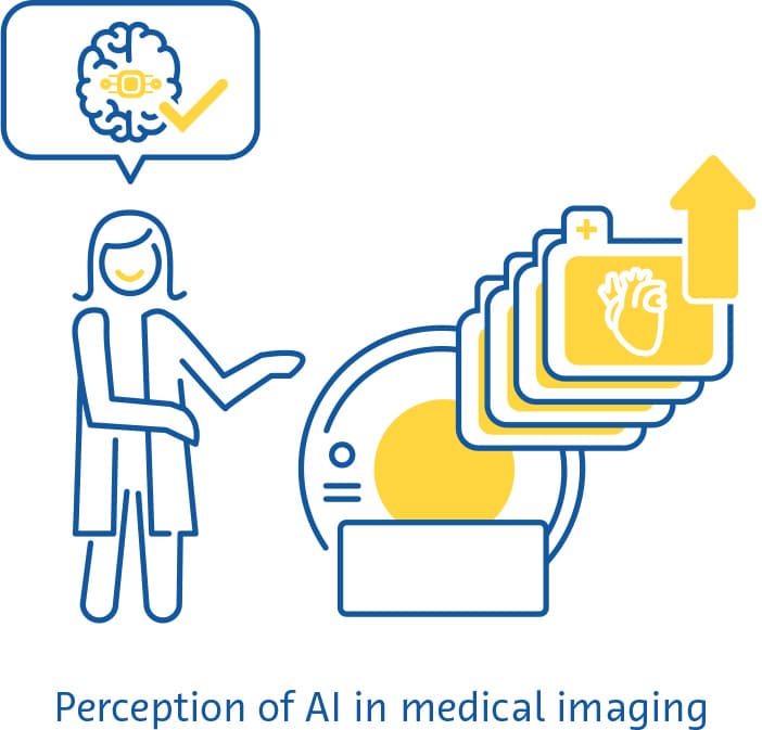
    Perception of AI

    The vast majority (77-84 %) of currently available AI solutions in medical imaging target CT, MRI and plain radiographs (Rezazade Mehrizi et al., 2021; van Leeuwen et al., 2021). Nuclear imaging techniques, such as positron emission tomography (PET). provide unique information not readily gained from other modalities. PET has thus far been largely neglected in terms of AI research (Rezazade Mehrizi et al., 2021; van Leeuwen et al., 2021), and is thus a potentially promising avenue for the field’s expansion.

    AI research is also expected to undergo a shift in the type of data being used. The typical inpatient receives more than one imaging study during their hospital stay (Shinagare et al., 2014). Despite this, only about 3 % of current AI-based radiology solutions combine data from multiple modalities (Rezazade Mehrizi et al., 2021; van Leeuwen et al., 2021). Combining data from multiple imaging sources may improve the diagnostic capabilities of AI solutions. Furthermore, future AI solutions in radiology are likely to combine imaging information, clinical information, as well as non- imaging diagnostic tests (Huang et al., 2020). By doing this, AI solutions may be able to identify patterns in the data collected during a patient’s hospital stay that may not be readily identifiable by healthcare workers (Rockenbach, 2021). This could ultimately lead to more accurate diagnoses and could help inform better and more personalized treatment decisions.

    The expectations for AI-based medical imaging solutions are also likely to shift from the current focus of triage, image enhancement and automation. With increasing algorithmic complexity, data availability, and experience with these tools, this shift may lead to AI solutions reaching specific diagnoses and recommending specific steps in a patient’s management plan. Similar to how the introduction of the first AI tools for image screening and processing around 2018 spurred investment in the field, marketing analyses predict a similar investment boost in the next few years as AI tools providing specific diagnoses and management steps become more widespread (Michoud et al., 2019).

    One important criticism of the current, arguably still nascent, landscape of AI in medical imaging is that it is too fragmented. Radiology professionals would likely welcome a more streamlined integration of AI solutions in their daily workflow. This includes seamless integration of these solutions into established radiology workflows, with as much as possible happening “in the background” without user input. Furthermore, the outputs of these solutions could be integrated into available radiological information systems. Consequently, the field could move from the plethora of currently available niche AI solutions, each targeted towards a single very specific application, to broader software suites that perform many different functions for a given imaging modality or body region.

    The fragmented investment in the AI in medical imaging market (Alexander et al., 2020) fosters innovation, allowing many players to test out different strategies in this emerging field. However, in the long term, consolidation may increase adoption and stimulate the kind of seamless integration into existing workflows that is needed, allowing fewer companies to offer these solutions at scale (Alexander et al., 2020).

    Challenges

    Quality and reporting of evidence

    In a review of 100 CE-marked AI solutions, 64 % of them had no peer-reviewed scientific evidence for their efficacy (van Leeuwen et al., 2021). Where there was scientific evidence, the level was low, rarely exceeding the demonstration of diagnostic accuracy (van Leeuwen et al., 2021). Another systematic review of the evidence for deep learning algorithms in medical imaging found a generally high diagnostic accuracy, albeit with a high risk of bias across studies (Aggarwal et al., 2021). The main sources of bias include the lack of external validation (D. W. Kim et al., 2019; Liu et al., 2019), insufficiently detailed reporting of results (Liu et al., 2019), retrospective study design (Nagendran et al., 2020), and the inaccessibility of data and code to reviewers and readers (Nagendran et al., 2020).

    Overall, studies on AI tools have shown a worrying lack of standardized reporting and adherence to recommended reporting guidelines (Aggarwal et al., 2021; Yusuf et al., 2020). This is despite the fact that several extensions to established reporting guidelines, as well as AI-specific guidelines, are currently available (Shelmerdine et al., 2021). Widespread implementation of these guidelines should be a focus of AI developers in the future.

    AI developers should also be cognizant that the currently “acceptable” level of evidence for AI-based solutions is likely to become obsolete in the near future. Both regulators and potential users will likely demand higher levels of evidence for these solutions, akin to the evidence required for new pharmaceutical drugs. In the next few years, will see more of these AI solutions being tested in randomized clinical trials. In the more distant future, it is plausible that such expectations will go beyond providing evidence of the safety, efficacy, or diagnostic performance of these solutions, to the demonstration that they provide added monetary or societal value.

    Rising up to the challenge of improving the quality and reporting of evidence for AI-based solutions may pay off in the long run. It could reduce the risk of bias in AI studies, could allow the thorough and transparent assessment of study quality by potential users and regulators, and could facilitate systematic reviews and meta-analyses. These steps may increase the trust in, and uptake of, AI-based solutions and ensure that they offer realistic, sustainable improvements in people’s lives.

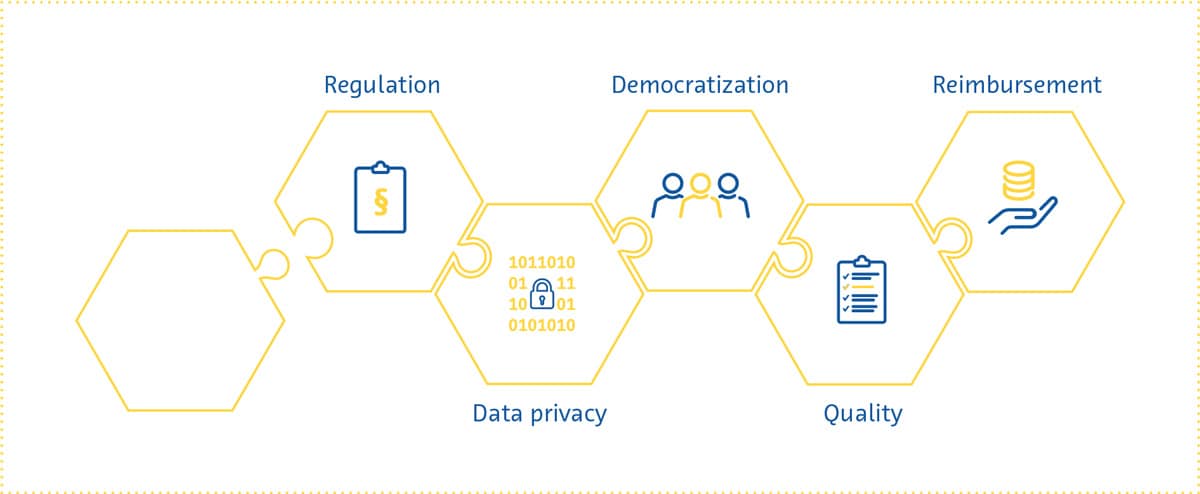
    Regulation

    Several aspects inherent to AI pose challenges to attempts at regulating it like other healthcare interventions. The inner workings of AI solutions are often opaque and difficult to comprehensively describe in a manner traditionally expected by regulatory bodies.

    The past few years have shown us that these regulatory challenges are far from intractable. Both the Food and Drug Administration and the European Commission have recently proposed initial regulatory frameworks for AI solutions (Center for Devices & Radiological Health, 2021; European Commission, 2021).

    In part as a response to the transparency necessary for regulatory approval, researchers have made substantial progress in making AI’s decision-making more understandable and explainable. This movement towards “interpretable AI” will gain further impetus in the near future as reliance on AI for real-world clinical decision-making increases.

    This has many advantages, including making regulatory approval easier, increasing trust in these solutions by users, minimizing biases, and improving the reproducibility of these solutions (Holzinger et al., 2017; Kolyshkina & Simoff, 2021; “Towards Trustable Machine Learning,” 2018; Yoon et al., 2021).

    Data privacy

    From development and testing to implementation, AI solutions in medical imaging require access to patient data. This has raised concerns about data privacy, which is a multifaceted and highly complex issue (Murdoch, 2021) that is prominently represented in the regulatory pathways of different countries (COCIR, the European Coordination Committee of the Radiological, Electromedical and Healthcare IT Industry, 2020). Suggested solutions to the data privacy question have ranged from those focusing on oversight to more technical approaches.

    The patients providing the data have to be made aware that they are doing so, as well as be informed about why and how their data will be used (Lotan et al., 2020), as explicitly stipulated in the EU’s General Data Protection Regulation (GDPR) (General Data Protection Regulation (GDPR) – Official Legal Text, 2016). Considering the fast-paced nature of the development of AI solutions, whether patients can be kept sufficiently informed as these algorithms are continuously retrained has been questioned (Kritikos, 2020). While fully anonymized data is not subject to such strict requirements under the GDPR (What Is Personal Data?, 2021), anonymization is exceedingly difficult to achieve for medical imaging data.

    The data privacy issue will have to be approached on several fronts. In addition to legislation governing the use of patient data, it is becoming increasingly clear that everyone involved in the development and use of AI solutions - developers, payers, regulatory bodies, researchers and radiologists - has a role to play in ensuring that the data is protected and used responsibly.

    Moreover, the next few years will likely see further research into technical approaches to strengthen data protection. These include better ways to reduce the chances of data being traced back to individuals, methods for keeping sensitive data stored locally even when the algorithm being trained is hosted in some “central” location, data perturbation to minimize the information within a given dataset pertaining to individual patients, and data encryption (G. Kaissis et al., 2021; G. A. Kaissis et al., 2020).

    hexagon


    Democratization

    If AI in medical imaging is to live up to its potential, the algorithms being developed have to work for everyone. This “democratization” of AI involves ensuring that healthcare providers have the knowledge and skills needed to use AI-based solutions. With a few exceptions (Paranjape et al., 2019), medical student curricula currently include little to no dedicated education about AI (Banerjee et al., 2021; Blease et al., 2022). Surveys from around the world have shown that medical students’ and doctors’ (Ahmed et al., 2022; Bisdas et al., 2021; Collado-Mesa et al., 2018; Kansal et al., 2022; Pinto Dos Santos et al., 2019; Sit et al., 2020) exposure to AI during training was low despite the high demand for more AI education (Kansal et al., 2022; Ooi et al., 2021; Sit et al., 2020). In addition, there are still large differences between genders and countries in the perceived knowledge about AI amongst medical students (Bisdas et al., 2021). There are many reasons for these differences and many challenges associated with the widespread integration of AI education into healthcare training curricula. In the coming years, strategies to tackle these issues should be investigated to ensure that future healthcare providers are equipped with the knowledge and skills they need to work in an environment where AI plays a growing role.

    Democratization also involves ensuring that patients of different genders, lifestyles, ethnicities, and geographical locations can benefit from AI-based solutions. For this to happen, these solutions have to be accessible and their performance generalizable. The latter requires the acquisition of diverse data from multiple institutions, preferably from multiple countries, for training AI-based solutions. It also requires the implementation of safeguards to ensure that sources of bias throughout the development process are not propagated to the trained algorithm (Vokinger et al., 2021), an issue that has only recently come to the forefront (Larrazabal et al., 2020; Obermeyer et al., 2019; Seyyed-Kalantari et al., 2021).

    Reimbursement

    As countries’ policies for regulating AI in healthcare gradually begin to take shape, one important aspect that needs attention is who will pay for these AI solutions, and according to what framework.

    Many consider Germany’s 2020 Digital Supply Act a step in the right direction for reimbursement of digital health solutions. Under this policy, digital applications prescribed by physicians are reimbursable by statutory health insurance if they are proven to be safe, be compliant with data privacy statutes, and improve patient care. The UK, on the other hand, has released a guide for potential buyers of AI-based solutions, which serves as a starting point for companies to prepare for reimbursement applications (A Buyer’s Guide to AI in Health and Care, 2020).

    Thus far, reimbursement success stories in the digital health space have been few and far between (Brink- mann-Sass et al., 2020; Hassan, 2021). This is in part due to requirements varying greatly by country (COCIR, the European Coordination Committee of the Radiological, Electromedical and Healthcare IT Industry, 2020). In general, providers of digital health solutions will need to provide evidence for the overall value that these solutions bring, including detailed health economics studies showing potential cost savings.

    Radiology’s position as a service provider to multiple hospital departments means that AI-based solutions in this space will be expected to show a far-reaching impact (van Duffelen, 2021). Companies will need to show short-term value (e.g. faster/better image reading and reporting) as well as long-term value (e.g. early diagnosis and treatment, disease prevention, reduction in unnecessary follow-up). The coming years will see companies compete to demonstrate such impact, while at the same time experimenting with different pricing models and navigating the evolving bureaucratic reimbursement landscape.

    Over the past few years, the field of AI in medical imaging has undergone a rapid but steady transformation. AI can now achieve things in radiology that few people thought possible a mere decade ago. The field is also gradually overcoming one of its most significant perceived hurdles - regulatory approval. In addition, while fear and scepticism dominated radiologists’ perception of the future of AI in their speciality a few years ago, this is no longer the case.

    The massive progress and interest in the field of AI in medical imaging is expected to continue into 2022 and beyond. Several exciting transformations await the field - it will likely expand its focus in the coming years to improve radiology workflow efficiency, involve hitherto neglected imaging modalities, combine data from multiple modalities, and provide more concrete diagnostic predictions and management recommendations. Easy-to-use and comprehensive software suites utilizing AI will be incorporated into existing radiology workflows, making radiologists’ and radiographers’ work easier and more efficient.

    As in any rapidly growing field, several scientific, regulatory, and economic challenges face AI in medical imaging. But the past few years have shown us that even the most difficult problems can be solved. Developers and users of AI-based solutions need to be aware of these issues so that they can adapt their strategies to changing expectations on a regulatory and societal level. Doing this will allow them to thrive in a fascinating field with the potential to improve virtually every aspect of healthcare.

    AAMC Report Reinforces Mounting Physician Shortage. (2021). AAMC. https://www.aamc.org/news-insights/press- releases/aamc-report-reinforces-mounting-physician-shortage

    A buyer’s guide to AI in health and care. (2020). NHS Transformation Directorate. https://www.nhsx.nhs.uk/ai-lab/ explore-all-resources/adopt-ai/a-buyers-guide-to-ai-in-health- and-care/

    Aggarwal, R., Sounderajah, V., Martin, G., Ting, D. S. W., Karthikesalingam, A., King, D., Ashrafian, H., & Darzi, A. (2021). Diagnostic accuracy of deep learning in medical imaging: a systematic review and meta-analysis. NPJ Digital Medicine, 4(1), 65.

    Ahmed, Z., Bhinder, K. K., Tariq, A., Tahir, M. J., Mehmood, Q., Tabassum, M. S., Malik, M., Aslam, S., Asghar, M. S., & Yousaf, Z. (2022). Knowledge, attitude, and practice of artificial intelligence among doctors and medical students in Pakistan: A cross-sectional online survey. Annals of Medicine and Surgery (2012), 76, 103493.

    AI Central. (n.d.). Retrieved February 23, 2022, from https://aicentral.acrdsi.org/

    AI for Radiology. (n.d.). Retrieved February 23, 2022, from https://grand-challenge.org/aiforradiology/

    Alexander, A., Jiang, A., Ferreira, C., & Zurkiya, D. (2020). An Intelligent Future for Medical Imaging: A Market Outlook on Artificial Intelligence for Medical Imaging. Journal of the American College of Radiology: JACR, 17(1 Pt B), 165–170.

    Allen, B., Agarwal, S., Coombs, L., Wald, C., & Dreyer, K. (2021). 2020 ACR Data Science Institute Artificial Intelligence Survey. Journal of the American College of Radiology: JACR, 18(8), 1153–1159.

    Banerjee, M., Chiew, D., Patel, K. T., Johns, I., Chappell, D., Linton, N., Cole, G. D., Francis, D. P., Szram, J., Ross, J., & Zaman, S. (2021). The impact of artificial intelligence on clinical education: perceptions of postgraduate trainee doctors in London (UK) and recommendations for trainers. BMC Medical Education, 21(1), 429.

    Bash, S., Johnson, B., Gibbs, W., Zhang, T., Shankaranarayanan, A., & Tanenbaum, L. N. (2021). Deep Learning Image Processing Enables 40 % Faster Spinal MR Scans Which Match or Exceed Quality of Standard of Care : A Prospective Multicenter Multireader Study. Clinical Neuroradiology. https://doi.org/10.1007/s00062-021-01121-2

    Bash, S., Wang, L., Airriess, C., Zaharchuk, G., Gong, E., Shankaranarayanan, A., & Tanenbaum, L. N. (2021). Deep Learning Enables 60 % Accelerated Volumetric Brain MRI While Preserving Quantitative Performance: A Prospective, Multicenter, Multireader Trial. AJNR. American Journal of Neuroradiology, 42(12), 2130–2137.

    Bennett, C. C., & Hauser, K. (2013). Artificial intelligence framework for simulating clinical decision-making: a Markov decision process approach. Artificial Intelligence in Medicine, 57(1), 9–19.

    Bisdas, S., Topriceanu, C.-C., Zakrzewska, Z., Irimia, A.-V., Shakallis, L., Subhash, J., Casapu, M.-M., Leon-Rojas, J., Pinto Dos Santos, D., Andrews, D. M., Zeicu, C., Bouhuwaish, A. M., Lestari, A. N., Abu-Ismail, L. ’i, Sadiq, A. S., Khamees, A. ’atasim, Mohammed, K. M. G., Williams, E., Omran, A. I.,… Ebrahim, E. H. (2021). Artificial Intelligence in Medicine: A Multinational Multi-Center Survey on the Medical and Dental Students’ Perception. Frontiers in Public Health, 9, 795284.

    Blease, C., Kharko, A., Bernstein, M., Bradley, C., Houston, M., Walsh, I., Hägglund, M., DesRoches, C., & Mandl, K. D. (2022). Machine learning in medical education: a survey of the experiences and opinions of medical students in Ireland. BMJ Health & Care Informatics, 29(1). https://doi.org/10.1136/ bmjhci-2021-100480

    Brown, A. D., & Marotta, T. R. (2018). Using machine learning for sequence-level automated MRI protocol selection in neuroradiology. Journal of the American Medical Informatics Association: JAMIA, 25(5), 568–571.

    Center for Devices, & Radiological Health. (2021, June 22). Digital Health Software Precertification (Pre-Cert) program.
    U.S. Food and Drug Administration. https://www.fda.gov/ medical-devices/digital-health-center-excellence/digital-health- software-precertification-pre-cert-program

    Clinical radiology UK workforce census 2019 report. (2019). https://www.rcr.ac.uk/publication/clinical-radiology-uk- workforce-census-2019-report

    COCIR, the European Coordination Committee of the Radiological, Electromedical and Healthcare IT Industry. (2020). Market Access Pathways for Digital Health Solutions. https://www.cocir.org/fileadmin/Publications_2020/20062_ COCIR_Market_Access_Pathways_Digital_Health.pdf

    Collado-Mesa, F., Alvarez, E., & Arheart, K. (2018). The Role of Artificial Intelligence in Diagnostic Radiology: A Survey at a Single Radiology Residency Training Program. Journal of the American College of Radiology: JACR, 15(12), 1753–1757.

    Core Health Indicators in the WHO European Region 2015. Special focus: Human resources for health. (2017, August 14). World Health Organization. https://www.euro.who.int/en/ data-and-evidence/evidence-resources/core-health-indicators- in-the-who-european-region/core-health-indicators-in-the- who-european-region-2015.-special-focus-human-resources- for-health

    Dantas, L. F., Fleck, J. L., Cyrino Oliveira, F. L., & Hamacher, S. (2018). No-shows in appointment scheduling - a systematic literature review. Health Policy, 122(4), 412–421.

    Esses, S. J., Lu, X., Zhao, T., Shanbhogue, K., Dane, B., Bruno, M., & Chandarana, H. (2018). Automated image quality evaluation of T2 -weighted liver MRI utilizing deep learning architecture. Journal of Magnetic Resonance Imaging: JMRI, 47(3), 723–728.

    European Commission. (2021). Proposal for a Regulation Of The European Parliament And Of The Council Laying Down
    Harmonised Rules On Artificial Intelligence (Artificial Intelligence Act) And Amending Certain Union Legislative Act.
    https://eur-lex. europa.eu/resource.html?uri=cellar:e0649735-a372-11eb- 9585-01aa75ed71a1.0001.02/DOC_1&format=PDF

    General Data Protection Regulation (GDPR) – Official Legal Text. (2016, July 13). General Data Protection Regulation (GDPR). https://gdpr-info.eu/

    Hata, A., Yanagawa, M., Yoshida, Y., Miyata, T., Tsubamoto, M., Honda, O., & Tomiyama, N. (2020). Combination of Deep Learning-Based Denoising and Iterative Reconstruction for Ultra-Low-Dose CT of the Chest: Image Quality and Lung-RADS Evaluation. AJR. American Journal of Roentgenology, 215(6), 1321–1328.

    Hauptmann, A., Arridge, S., Lucka, F., Muthurangu, V., & Steeden, J. A. (2019). Real-time cardiovascular MR with
    spatio-temporal artifact suppression using deep learning-proof of concept in congenital heart disease. Magnetic Resonance in Medicine: Official Journal of the Society of Magnetic Resonance in Medicine / Society of Magnetic Resonance in Medicine, 81(2), 1143–1156.

    Higaki, T., Nakamura, Y., Zhou, J., Yu, Z., Nemoto, T., Tatsugami, F., & Awai, K. (2020). Deep Learning Reconstruction at CT: Phantom Study of the Image Characteristics. Academic Radiology, 27(1), 82–87.

    Holzinger, A., Biemann, C., Pattichis, C. S., & Kell, D. B. (2017). What do we need to build explainable AI systems for the medical domain? In arXiv [cs.AI]. arXiv. http://arxiv.org/ abs/1712.09923

    Hötker, A. M., Da Mutten, R., Tiessen, A., Konukoglu, E., & Donati, O. F. (2021). Improving workflow in prostate MRI: AI- based decision-making on biparametric or multiparametric MRI. Insights into Imaging, 12(1), 112.

    Huang, S.-C., Pareek, A., Seyyedi, S., Banerjee, I., & Lungren, M. P. (2020). Fusion of medical imaging and electronic health records using deep learning: a systematic review and implementation guidelines. NPJ Digital Medicine, 3, 136.

    Kaissis, G. A., Makowski, M. R., Rückert, D., & Braren, R. F. (2020). Secure, privacy-preserving and federated machine learning in medical imaging. Nature Machine Intelligence, 2(6), 305–311.

    Kaissis, G., Ziller, A., Passerat-Palmbach, J., Ryffel, T., Usynin, D., Trask, A., Lima, I., Mancuso, J., Jungmann, F., Steinborn, M.-M., Saleh, A., Makowski, M., Rueckert, D., & Braren, R. (2021). End-to-end privacy preserving deep learning on multi-institutional medical imaging. Nature Machine Intelligence, 3(6), 473–484.

    Kansal, R., Bawa, A., Bansal, A., Trehan, S., Goyal, K., Goyal, N., & Malhotra, K. (2022). Differences in Knowledge and Perspectives on the Usage of Artificial Intelligence Among Doctors and Medical Students of a Developing Country: A Cross- Sectional Study. Cureus, 14(1), e21434.

    Katsari, K., Penna, D., Arena, V., Polverari, G., Ianniello, A., Italiano, D., Milani, R., Roncacci, A., Illing, R. O., & Pelosi, E. (2021). Artificial intelligence for reduced dose 18F-FDG PET examinations: a real-world deployment through a standardized framework and business case assessment. EJNMMI Physics, 8(1), 25.

    Kerpel, A., Marom, E. M., Green, M., Eifer, M., Konen, E., Mayer, A., & Betancourt Cuellar, S. L. (2021). Ultra-Low Dose Chest CT with Denoising for Lung Nodule Detection. The Israel Medical Association Journal: IMAJ, 23(9), 550–555.

    Kim, D. W., Jang, H. Y., Kim, K. W., Shin, Y., & Park, S. H. (2019).
    Design Characteristics of Studies Reporting the Performance of Artificial Intelligence Algorithms for Diagnostic Analysis of
    Medical Images: Results from Recently Published Papers. Korean Journal of Radiology: Official Journal of the Korean Radiological Society, 20(3), 405–410.

    Kim, K. H., & Park, S.-H. (2017). Artificial neural network for suppression of banding artifacts in balanced steady-state free precession MRI. Magnetic Resonance Imaging, 37, 139–146.

    Kolyshkina, I., & Simoff, S. (2021). Interpretability of Machine Learning Solutions in Public Healthcare: The CRISP-ML Approach. Frontiers in Big Data, 4, 660206.

    Komorowski, M., Celi, L. A., Badawi, O., Gordon, A. C., & Faisal, A. A. (2018). The Artificial Intelligence Clinician learns optimal treatment strategies for sepsis in intensive care. Nature Medicine, 24(11), 1716–1720.

    Kritikos, M. (2020). What if artificial intelligence in medical imaging could accelerate Covid-19 treatment? Think Tank
    - European Parliament. https://www.europarl.europa.eu/ thinktank/en/document/EPRS_ATA(2020)656333

    Kurasawa, H., Hayashi, K., Fujino, A., Takasugi, K., Haga, T., Waki, K., Noguchi, T., & Ohe, K. (2016). Machine-Learning- Based Prediction of a Missed Scheduled Clinical Appointment by Patients With Diabetes. Journal of Diabetes Science and Technology, 10(3), 730–736.

    Larrazabal, A. J., Nieto, N., Peterson, V., Milone, D. H., & Ferrante, E. (2020). Gender imbalance in medical imaging datasets produces biased classifiers for computer-aided diagnosis. Proceedings of the National Academy of Sciences of the United States of America, 117(23), 12592–12594.

    Larson, D. B., Johnson, L. W., Schnell, B. M., Salisbury, S. R., & Forman, H. P. (2011). National trends in CT use in the emergency department: 1995-2007. Radiology, 258(1), 164–173.

    Le, V., Frye, S., Botkin, C., Christopher, K., Gulaka, P., Sterkel, B., Frye, R., Muzaffar, R., & Osman, M. (2020). Effect of PET Scan with Count Reduction Using AI-Based Processing Techniques on Image Quality. Journal of Nuclear Medicine: Official Publication, Society of Nuclear Medicine, 61(supplement 1), 3095–3095.

    Levin, D. C., Parker, L., & Rao, V. M. (2017). Recent Trends in Imaging Use in Hospital Settings: Implications for Future Planning. Journal of the American College of Radiology: JACR, 14(3), 331–336.

    Liu, X., Faes, L., Kale, A. U., Wagner, S. K., Fu, D. J., Bruynseels, A., Mahendiran, T., Moraes, G., Shamdas, M., Kern, C., Ledsam, J. R., Schmid, M. K., Balaskas, K., Topol, E.J., Bachmann, L. M., Keane, P. A., & Denniston, A. K. (2019). A comparison of deep learning performance against health-care professionals in detecting diseases from medical imaging: a systematic review and meta-analysis. The Lancet. Digital Health, 1(6), e271–e297.

    Lotan, E., Tschider, C., Sodickson, D. K., Caplan, A. L., Bruno, M., Zhang, B., & Lui, Y. W. (2020). Medical Imaging and Privacy in the Era of Artificial Intelligence: Myth, Fallacy, and the Future. Journal of the American College of Radiology: JACR, 17(9), 1159–1162.

    Mairhöfer, D., Laufer, M., Simon, P. M., Sieren, M., Bischof, A., Käster, T., Barth, E., Barkhausen, J., & Martinetz, T. (2021). An AI-based Framework for Diagnostic Quality Assessment of Ankle Radiographs. https://openreview.net/pdf?id=bj04hJss_xZ

    Makeeva, V., Gichoya, J., Hawkins, C. M., Towbin, A. J., Heilbrun, M., & Prater, A. (2019). The Application of Machine Learning to Quality Improvement Through the Lens of the Radiology Value Network. Journal of the American College of Radiology: JACR, 16(9 Pt B), 1254–1258.

    McLeavy, C. M., Chunara, M. H., Gravell, R. J., Rauf, A., Cushnie, A., Staley Talbot, C., & Hawkins, R. M. (2021). The future of CT: deep learning reconstruction. Clinical Radiology, 76(6), 407–415.

    Medical AI Evaluation. (n.d.). Retrieved February 23, 2022, from https://ericwu09.github.io/medical-ai-evaluation/

    Michoud, L., Tschudi, Y., & Villien, Y. (2019). Artificial Intelligence for Medical Imaging: Market and Technology Report 2020. Yole Développement. https://s3.i-micronews.com/ uploads/2020/01/YDR20059-AI-for-Medical-Imaging_Yole_ sample.pdf

    Murdoch, B. (2021). Privacy and artificial intelligence: challenges for protecting health information in a new era. BMC Medical Ethics, 22(1), 122.

    Nagendran, M., Chen, Y., Lovejoy, C. A., Gordon, A. C., Komorowski, M., Harvey, H., Topol, E. J., Ioannidis, J. P. A., Collins, G. S., & Maruthappu, M. (2020). Artificial intelligence versus clinicians: systematic review of design, reporting standards, and claims of deep learning studies. BMJ, 368. https://doi.org/10.1136/bmj.m689

    Nelson, A., Herron, D., Rees, G., & Nachev, P. (2019). Predicting scheduled hospital attendance with artificial intelligence. Npj Digital Medicine, 2(1), 26.

    Obermeyer, Z., Powers, B., Vogeli, C., & Mullainathan, S. (2019). Dissecting racial bias in an algorithm used to manage the health of populations. Science, 366(6464), 447–453.

    Ooi, S. K. G., Makmur, A., Soon, A. Y. Q., Fook-Chong, S., Liew, C., Sia, S. Y., Ting, Y. H., & Lim, C. Y. (2021). Attitudes toward artificial intelligence in radiology with learner needs assessment within radiology residency programmes: a national multi-programme survey. Singapore Medical Journal, 62(3), 126–134.

    Paranjape, K., Schinkel, M., Nannan Panday, R., Car, J., & Nanayakkara, P. (2019). Introducing Artificial Intelligence Training in Medical Education. JMIR Medical Education, 5(2), e16048.

    Park, J., Hwang, D., Kim, K. Y., Kang, S. K., Kim, Y. K., & Lee, J. S. (2018). Computed tomography super-resolution using deep convolutional neural network. Physics in Medicine and Biology, 63(14), 145011.

    Pinto Dos Santos, D., Giese, D., Brodehl, S., Chon, S. H., Staab, W., Kleinert, R., Maintz, D., & Baeßler, B. (2019). Medical students’ attitude towards artificial intelligence: a multicentre survey. European Radiology, 29(4), 1640–1646.

    Population ages 65 and above. (n.d.). The World Bank. Retrieved February 23, 2022, from https://data.worldbank.org/ indicator/SP.POP.65UP.TO.ZS

    Prevedello, L. M., Erdal, B. S., Ryu, J. L., Little, K. J., Demirer, M., Qian, S., & White, R. D. (2017). Automated Critical Test Findings Identification and Online Notification System Using Artificial Intelligence in Imaging. Radiology, 285(3), 923–931.

    Rezazade Mehrizi, M. H., van Ooijen, P., & Homan, M. (2021). Applications of artificial intelligence (AI) in diagnostic radiology: a technography study. European Radiology, 31(4), 1805–1811.

    Rockenbach, M. A. B. (2021, June 13). Multimodal AI in healthcare: Closing the gaps. CodeX. https://medium.com/codex/ multimodal-ai-in-healthcare-1f5152e83be2

    Rudie, J. D., Gleason, T., Barkovich, M. J., Wilson, D. M., Shankaranarayanan, A., Zhang, T., Wang, L., Gong, E., Zaharchuk, G., & Villanueva-Meyer, J. E. (2022). Clinical Assessment of Deep Learning–based Super-Resolution for 3D Volumetric Brain MRI. Radiology: Artificial Intelligence, e210059.

    Schreiber-Zinaman, J., & Rosenkrantz, A. B. (2017). Frequency and reasons for extra sequences in clinical abdominal MRI examinations. Abdominal Radiology (New York), 42(1), 306–311.

    Seyyed-Kalantari, L., Zhang, H., McDermott, M. B. A., Chen, I. Y., & Ghassemi, M. (2021). Underdiagnosis bias of artificial intelligence algorithms applied to chest radiographs in under-served patient populations. Nature Medicine, 27(12), 2176–2182.

    Shelmerdine, S. C., Arthurs, O. J., Denniston, A., & Sebire, N. J. (2021). Review of study reporting guidelines for clinical studies using artificial intelligence in healthcare. BMJ Health & Care Informatics, 28(1). https://doi.org/10.1136/ bmjhci-2021-100385

    Shinagare, A. B., Ip, I. K., Abbett, S. K., Hanson, R., Seltzer, S. E., & Khorasani, R. (2014). Inpatient imaging utilization: trends of the past decade. AJR. American Journal of Roentgenology, 202(3), W277–W283.

    Singh, R., Digumarthy, S. R., Muse, V. V., Kambadakone, A. R., Blake, M. A., Tabari, A., Hoi, Y., Akino, N., Angel, E., Madan, R., & Kalra, M. K. (2020). Image Quality and Lesion Detection on Deep Learning Reconstruction and Iterative Reconstruction of Submillisievert Chest and Abdominal CT. AJR. American Journal of Roentgenology, 214(3), 566–573.

    Sit, C., Srinivasan, R., Amlani, A., Muthuswamy, K., Azam, A., Monzon, L., & Poon, D. S. (2020). Attitudes and perceptions of UK medical students towards artificial intelligence and radiology: a multicentre survey. Insights into Imaging, 11(1), 14.

    Smith-Bindman, R., Miglioretti, D. L., Johnson, E., Lee, C., Feigelson, H. S., Flynn, M., Greenlee, R. T., Kruger, R. L., Hornbrook, M. C., Roblin, D., Solberg, L. I., Vanneman, N., Weinmann, S., & Williams, A. E. (2012). Use of diagnostic imaging studies and associated radiation exposure for patients enrolled in large integrated health care systems, 1996-2010. JAMA: The Journal of the American Medical Association, 307(22), 2400–2409.

    Smith-Bindman, R., Miglioretti, D. L., & Larson, E. B. (2008). Rising use of diagnostic medical imaging in a large integrated health system. Health Affairs, 27(6), 1491–1502.

    The Medical Futurist. (n.d.). The Medical Futurist. Retrieved February 23, 2022, from https://medicalfuturist.com/fda- approved-ai-based-algorithms/

    Towards trustable machine learning. (2018). Nature Biomedical Engineering, 2(10), 709–710.

    Trivedi, H., Mesterhazy, J., Laguna, B., Vu, T., & Sohn, J. H. (2018). Automatic Determination of the Need for Intravenous Contrast in Musculoskeletal MRI Examinations Using IBM Watson’s Natural Language Processing Algorithm. Journal of Digital Imaging, 31(2), 245–251.

    Tsao, D. N. (2020, July 27). AI in medical diagnostics 2020- 2030: Image recognition, players, clinical applications, forecasts: IDTechEx. https://www.idtechex.com/en/research- report/ai-in-medical-diagnostics-2020-2030-image- recognition-players-clinical-applications-forecasts/766

    van Duffelen, J. (2021, February 22). Making a case for buying medical imaging AI: How to define the return on investment.
    Aidence. https://www.aidence.com/articles/medical-imaging- ai-roi/

    van Leeuwen, K. G., Schalekamp, S., Rutten, M. J. C. M., van Ginneken, B., & de Rooij, M. (2021). Artificial intelligence in radiology: 100 commercially available products and their scientific evidence. European Radiology, 31(6), 3797–3804.

    Vokinger, K. N., Feuerriegel, S., & Kesselheim, A. S. (2021). Mitigating bias in machine learning for medicine. Communication & Medicine, 1, 25.

    Wang, S., Cao, G., Wang, Y., Liao, S., Wang, Q., Shi, J., Li, C., & Shen, D. (2021). Review and Prospect: Artificial Intelligence in Advanced Medical Imaging. Frontiers in Radiology, 1. https://doi. org/10.3389/fradi.2021.781868

    What is personal data? (2021, January 1). ICO - Information Commissioner’s Office; ICO. https://ico.org.uk/for-organisations/ guide-to-data-protection/guide-to-the-general-data-protection- regulation-gdpr/what-is-personal-data/what-is-personal-data

    WHO. (n.d.-a). Ageing and health. Retrieved February 23, 2022, from https://www.who.int/news-room/fact-sheets/detail/ ageing-and-health

    WHO. (n.d.-b). Noncommunicable diseases. Retrieved March 6, 2022, from https://www.who.int/news-room/fact-sheets/detail/ noncommunicable-diseases

    Winder, M., Owczarek, A. J., Chudek, J., Pilch-Kowalczyk, J., & Baron, J. (2021). Are We Overdoing It? Changes in Diagnostic Imaging Workload during the Years 2010-2020 including the Impact of the SARS-CoV-2 Pandemic. Healthcare (Basel, Switzerland), 9(11). https://doi.org/10.3390/healthcare9111557

    Winkel, D. J., Heye, T., Weikert, T. J., Boll, D. T., & Stieltjes, B. (2019). Evaluation of an AI-Based Detection Software for Acute Findings in Abdominal Computed Tomography Scans: Toward an Automated Work List Prioritization of Routine CT Examinations. Investigative Radiology, 54(1), 55–59.

    Wong, T. T., Kazam, J. K., & Rasiej, M. J. (2019). Effect of Analytics-Driven Worklists on Musculoskeletal MRI
    Interpretation Times in an Academic Setting. AJR. American Journal of Roentgenology, 1–5.

    Xu, F., Pan, B., Zhu, X., Gulaka, P., Xiang, L., Gong, E., Zhang, T., Wang, J., Lin, L., Ma, Y., & Gong, N.-J. (2020). Evaluation of Deep Learning Based PET Image Enhancement Method in Diagnosis of Lymphoma. Journal of Nuclear Medicine: Official Publication, Society of Nuclear Medicine, 61(supplement 1), 431–431.

    Yoon, C. H., Torrance, R., & Scheinerman, N. (2021). Machine learning in medicine: should the pursuit of enhanced interpretability be abandoned? Journal of Medical Ethics. https://doi.org/10.1136/medethics-2020-107102

    Yusuf, M., Atal, I., Li, J., Smith, P., Ravaud, P., Fergie, M., Callaghan, M., & Selfe, J. (2020). Reporting quality of studies using machine learning models for medical diagnosis: a systematic review. BMJ Open, 10(3), e034568.

    Zhang, D., Mishra, S., Brynjolfsson, E., Etchemendy, J., Ganguli, D., Grosz, B., Lyons, T., Manyika, J., Niebles, J. C., Sellitto, M., Shoham, Y., Clark, J., & Perrault, R. (2021). The AI Index 2021 Annual Report. In arXiv [cs.AI]. arXiv. http://arxiv.org/ abs/2103.06312

    Zhang, Y., & Yu, H. (2018). Convolutional Neural Network Based Metal Artifact Reduction in X-Ray Computed Tomography. IEEE Transactions on Medical Imaging, 37(6), 1370–1381.

    Zhu, B., Liu, J. Z., Cauley, S. F., Rosen, B. R., & Rosen, M. S. (2018). Image reconstruction by domain-transform manifold learning. Nature, 555(7697), 487–492.