Explore Your Local Site

Looks like you've landed on our   site. Let's take you home:    

Please note that the content and products on the    site might not be available in your region.


Choose the language:

  Homepage
Continue on the current website:  


Le rôle actuel et futur de l’intelligence artificielle dans le diagnostic et le dépistage du cancer du sein

Cancer du sein

Le cancer du sein est le type de cancer spécifique le plus répandu chez les femmes dans le monde (Sung et. al., 2021). Chez les femmes, le cancer du sein représente 1 cas de cancer sur 4 et 1 décès par cancer sur 6, se classant au premier rang en termes d'incidence dans la grande majorité des pays (159 pays sur 185) et de mortalité dans 110 pays (Sung et. al., 2021). La plupart des cas surviennent chez les femmes âgées de plus de 50 ans, mais ils peuvent également toucher des femmes plus jeunes. D'autres facteurs de risque comprennent une prédisposition génétique, des antécédents familiaux, l'apparition précoce des règles, un traitement hormonal substitutif, la consommation d'alcool et l'obésité (Łukasiewicz et al., 2021). 

Le sein est composé de lobules producteurs de lait, d'un système de canaux de transport et de tissu adipeux (Bazira et al., 2021). Tous les cancers du sein proviennent des cellules tapissant les unités lobulaires des canaux terminaux (l'unité fonctionnelle du sein) des canaux collecteurs. Le type de cancer du sein masculin le plus courant est le carcinome canalaire invasif, qui prend naissance dans les canaux galactophores et envahit les tissus voisins (Harbeck et al., 2019). Le développement du cancer du sein implique des mutations génétiques qui provoquent une prolifération cellulaire incontrôlée ainsi que les gènes BRCA1 et BRCA2, impliqués dans la réparation de l'ADN (Harbeck et al., 2019). Les récepteurs des oestrogènes et de la progestérone jouent un rôle important dans la physiopathologie ; tous les patients atteints de tumeurs exprimant ces récepteurs devraient recevoir un traitement hormonal pour bloquer l'activité des récepteurs des oestrogènes (Harbeck et al., 2019).

Le cancer du sein peut se manifester de différentes manières. Les caractéristiques cliniques les plus courantes sont la présence d’une masse dans le sein, des modifications de la taille du mamelon, un écoulement du mamelon et des modifications cutanées ainsi qu'une infection et/ou une inflammation du sein (Koo et al., 2017). Le cancer du sein à un stade précoce est souvent asymptomatique, ce qui souligne l'importance du dépistage systématique (Kalager et al., 2010).

Le cancer du sein est généralement diagnostiqué lors d’un dépistage ou en présence d’un symptôme (douleur ou masse palpable) qui déclenche un examen diagnostique (McDonald et al., 2016). Celui-ci est complété par des techniques d’imagerie permettant de rechercher des anomalies et de les caractériser plus en détail (McDonald et al., 2016). Une biopsie mammaire est généralement réalisée pour confirmer la présence d'un cancer en cas de suspicion, et permet également de déterminer son type spécifique si la lésion est cancéreuse (McDonald et al., 2016). Le cancer du sein est classé en fonction de l'étendue de la tumeur, de la propagation aux ganglions lymphatiques voisins, de la propagation à des sites distants, du statut des récepteurs aux oestrogènes, du statut des récepteurs de la progestérone, du statut HER2 et du grade du cancer (McDonald et al., 2016).

Il existe différents types de cancer du sein, et le traitement peut varier en fonction des caractéristiques moléculaires de la maladie, du stade, du type de cancer et du statut des récepteurs du patient (Hong et Xu, 2022). Le traitement implique généralement une association de différentes modalités et une équipe multidisciplinaire de professionnels de la santé (Hong et Xu, 2022). Les options chirurgicales vont des procédures conservatrices du sein à la mastectomie, où le sein entier est retiré (Hong et Xu, 2022). L'ablation des ganglions lymphatiques peut également s’avérer nécessaire pour évaluer l'étendue de la propagation du cancer (Hong et Xu, 2022). La radiothérapie est souvent utilisée après un traitement conservateur du sein ou une mastectomie (avec facteurs de risque) (Hong et Xu, 2022). La chimiothérapie systémique peut être administrée avant ou après la chirurgie, selon la situation spécifique (Hong et Xu, 2022). Les cancers du sein à récepteurs hormonaux positifs peuvent être traités par des médicaments qui bloquent les effets des oestrogènes et de la progestérone. L'immunothérapie est une option thérapeutique émergente pour certains cancers du sein, aidant le système immunitaire à reconnaître et à attaquer les cellules cancéreuses (Hong et Xu, 2022).
 

Techniques d'imagerie

Mammographie numérique

La mammographie numérique est la technique la plus souvent utilisée pour le dépistage du cancer du sein. Il s'agit d'une technique de sommation bidimensionnelle dans un tube à rayons X émet des rayons X, qui sont absorbés à différents degrés par les tissus et mesurés par un détecteur situé à l'autre extrémité. Les tissus plus denses apparaissent plus clairs sur les images résultantes que les tissus moins denses. Les seins sont comprimés pendant l'acquisition de l'image pour étendre le tissu mammaire sur une plus grande surface (Ikeda, 2011a). Cette technique permet de réduire le chevauchement entre les différents composants du tissu mammaire, de diminuer la diffusion des rayons X qui passent et d’améliorer le contraste. Deux vues de chaque sein sont généralement acquises : crânio-caudale (CC) et médio-latérale (MLO) (Ikeda, 2011a).
mammography

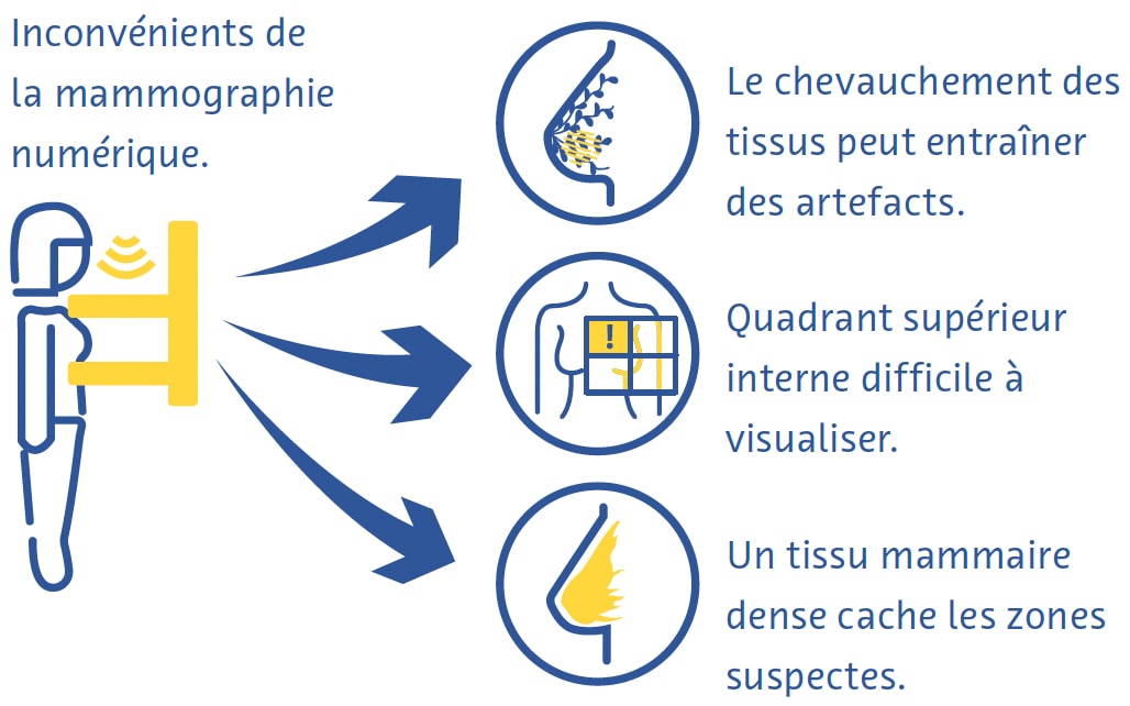
La mammographie numérique est une technique rapide et utile pour le dépistage du cancer du sein, mais elle présente néanmoins des inconvénients (Ikeda, 2011a). La compression mammaire peut être douloureuse et, malgré la compression, le chevauchement de différents tissus conduit souvent à des artefacts (Ikeda, 2011a). Le quadrant supérieur interne du sein, moins mobile car fixé à la paroi thoracique, est particulièrement difficile à visualiser en mammographie (Ikeda, 2011a). Le cancer peut également être très difficile à détecter à la mammographie dans les seins comportant une forte proportion de tissus denses (Ikeda, 2011a).

Tomosynthèse mammaire numérique

La tomosynthèse mammaire numérique (TMN) implique l'acquisition d'images à l'aide d'une source de rayons X qui se déplace le long d'un arc d'excursion. De fines tranches sont reconstruites, permettant des capacités d'imagerie 3D destinées à limiter autant que possible l'influence du tissu mammaire qui se chevauche. Cette méthode est particulièrement utile pour l'imagerie des lésions mammaires situées dans un parenchyme mammaire hétérogène et dense. Une étude a révélé que la TMN est plus sensible pour la détection du cancer du sein que la mammographie numérique (MN). La TMN peut être combinée avec la MN et une étude a révélé que l'utilisation d'une combinaison de ces techniques améliore la détection du cancer du sein (Alabousi et al., 2020 ; Lei et al., 2014 ; Skaane et al., 2019) et peut être combinée avec la mammographie. Cependant, la TMN prend plus de temps à acquérir que la mammographie et souffre d’artefacts de mouvements et autres (Tirada et al., 2019).

Échographie

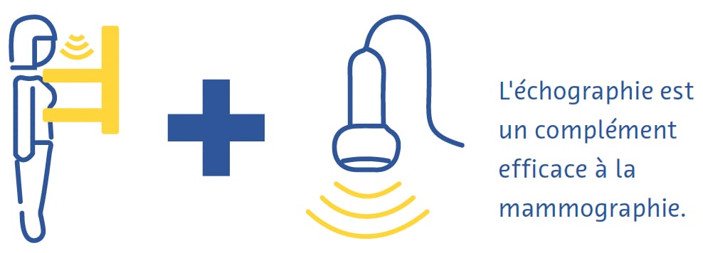
Dans l’échographie diagnostique, un transducteur émet des ondes sonores à haute fréquence qui traversent les tissus, rebondissent sur eux et créent des « échos » qui sont réfléchis et détectés par le transducteur. Ces échos sont ensuite traités pour créer des images en temps réel sur un moniteur en fonction du temps nécessaire aux échos pour se déplacer vers les tissus et revenir. Il s’agit d’une technique sûre et relativement peu coûteuse qui est souvent utilisée en complément de la mammographie (Ikeda, 2011b), notamment pour évaluer plus en détail un résultat palpable ou mammographique.
echographie

Elle peut même être utilisée comme modalité de dépistage primaire chez les femmes de moins de 30 ans ou chez les femmes enceintes ou allaitantes (Dixon, 2008 ; Ikeda, 2011b). L'échographie est très utile pour déterminer si une masse est kystique ou solide, quels types de marges elle présente et sa vascularisation (Dixon, 2008 ; Ikeda, 2011b). Elle permet également de détecter d'autres masses et ganglions lymphatiques axillaires suspects (Dixon, 2008 ; Ikeda, 2011b). Son principal inconvénient est que la qualité de l’examen dépend fortement de l’opérateur (Dixon, 2008 ; Ikeda, 2011b). 

Imagerie par résonance magnétique

À l’aide d’un puissant champ magnétique et d’une série d’ondes radiofréquences, l’imagerie par résonance magnétique (IRM) perturbe les noyaux d’hydrogène dans les tissus pour créer des images transversales détaillées du corps (Daniel et Ikeda, 2011 ; Mann et al., 2019). Étant donné que les tissus de compositions différentes réagissent de différentes manières à cette perturbation, l’IRM peut très bien détecter même des différences subtiles entre les types de tissus mous et est considérée comme la modalité la plus sensible pour diagnostiquer le cancer du sein (Daniel et Ikeda, 2011 ; Mann et al., 2019). Elle est principalement utilisée pour dépister les patients à haut risque selon les facteurs de risque génétiques ou acquis (Daniel et Ikeda, 2011).
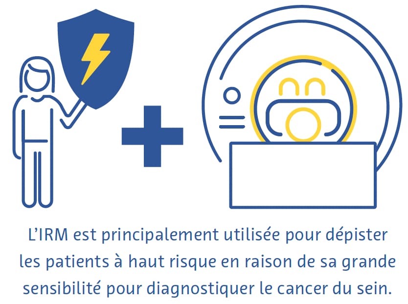
magnetique

L’IRM mammaire nécessite des antennes mammaires dédiées qui transmettent les ondes radiofréquences et reçoivent le signal généré. Les images sont souvent acquises avec une résolution spatiale dans le plan de 1 mm, une épaisseur de coupe inférieure à 3 mm et une suppression du signal provenant des lipides. Les séquences couramment utilisées comprennent les images pondérées en T2, l'imagerie pondérée en diffusion et l'IRM dynamique avec contraste amélioré. Pour réduire les faux positifs dus à des modifications non spécifiques du parenchyme mammaire, il est préférable de l'effectuer entre le 7e et le 13e jour du cycle menstruel (Daniel et Ikeda, 2011). Contrairement à la mammographie, l'IRM n'utilise pas de rayonnements ionisants et produit des images tridimensionnelles qui facilitent la détection de très petites lésions (DeMartini et Lehman, 2008 ; Shahid et al., 2016). L'IRM permet également une évaluation plus détaillée de la paroi thoracique que la mammographie et l'échographie (DeMartini et Lehman, 2008). Les inconvénients de l'IRM mammaire comprennent une faible sensibilité aux micro-calcifications, un coût élevé et le fait qu'elle soit contre-indiquée chez les personnes portant certains implants métalliques (Daniel et Ikeda, 2011).  
 

Défis du dépistage et du diagnostic

Malgré les preuves du bénéfice global du dépistage du cancer du sein (Dibden et al., 2020 ; Kalager et al., 2010 ; Tabár et al., 2019), ce dernier souffre de plusieurs défis techniques et logistiques. Plus de la moitié des femmes dépistées chaque année pendant 10 ans auront un test faussement positif (Hubbard et al., 2011). Cela a des conséquences vastes et significatives, notamment le fardeau physique et émotionnel des biopsies inutiles et l'augmentation des coûts des soins de santé (Nelson, Pappas et al., 2016 ; Ong et Mandl, 2015). Le cancer du sein passe souvent inaperçu lors du dépistage, en particulier chez les femmes aux seins denses (Banks et al., 2006).
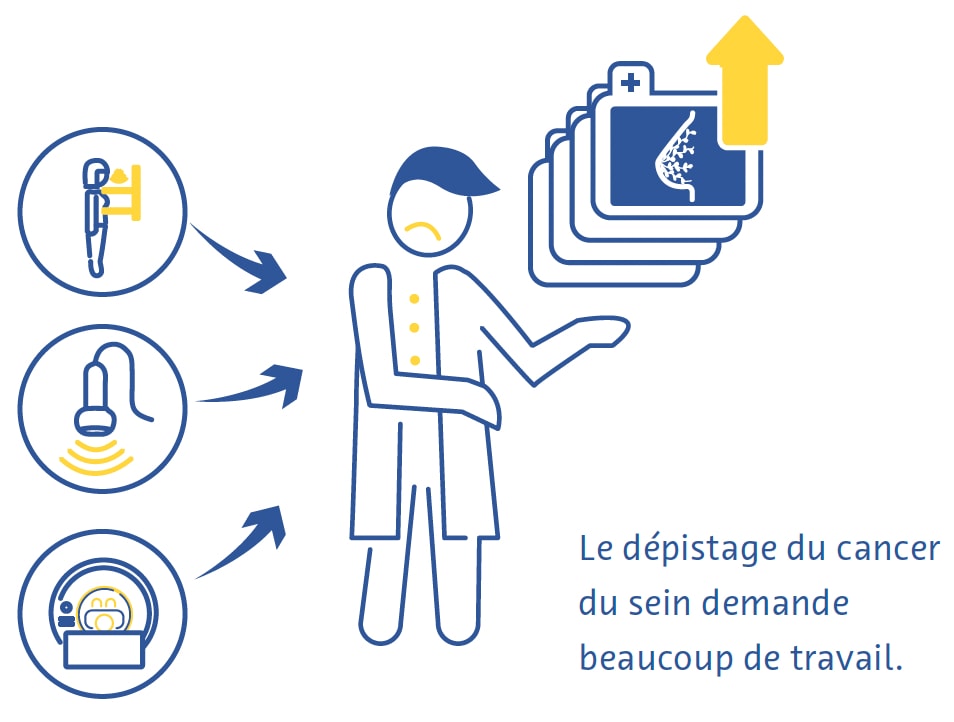
diagnostic

Le dépistage du cancer du sein nécessite du personnel hautement qualifié, notamment des radiologues et des radiographes, dont il existe actuellement une pénurie mondiale (Moran et Warren-Forward, 2012 ; Rimmer, 2017 ; Wing et Langelier, 2009). Ce problème est aggravé par le fait que la norme de soins en matière de dépistage du cancer du sein dans de nombreux pays européens est que chaque examen est lu indépendamment par deux radiologues (Giordano et al., 2012) et que, dans certains pays, comme les États-Unis, les obstacles à l'obtention de l'autorisation d'interpréter des mammographies sont élevés en raison des normes strictes de certification professionnelle (Food and Drug Administration, 2001).

Il existe également des obstacles importants à l’adoption du dépistage du cancer du sein dans le monde. Ceux-ci incluent le manque ou l’accès difficile aux programmes de dépistage, le manque de connaissances ou l’incompréhension des avantages de ces programmes, ainsi que les obstacles sociaux et culturels (Mascara et Constantinou, 2021).
 

Rôle de l'intelligence artificielle

Améliorations techniques

Jusqu’à présent, peu d’études publiées concernaient directement l’utilisation de l’IA pour apporter des améliorations techniques aux examens des seins. Une application disponible dans le commerce fournit aux radiologues un retour d'information en temps réel sur l'adéquation du positionnement du patient sur les mammographies. (Volpara Health, 2022). D'autres applications de l'IA se sont concentrées sur la réduction des doses de rayonnement (J. Liu et al., 2018), l'amélioration de la reconstruction des images (Kim et al., 2016) et la réduction du bruit et des artefacts sur la TMN (Garrett et al., 2018).

La TMN est fréquemment associée à la mammographie numérique pour le dépistage du cancer du sein, qui double la dose de rayonnement reçue par les patients (Svahn et al., 2015). Pour éviter cela, la génération de mammographies synthétiques à partir de données issues de la TMN suscite un intérêt croissant (Chikarmane et al., 2023). Dans une vaste étude prospective norvégienne, les précisions de la TMN combinée à la mammographie numérique ou à la mammographie synthétique pour la détection du cancer du sein étaient très similaires (Skaane et al., 2019). Des études récentes ont cherché à améliorer la qualité de la mammographie synthétique grâce à l’IA, avec des résultats prometteurs (Balleyguier et al., 2017 ; James et al., 2018). 

Améliorations du diagnostic

Évaluation de la densité mammaire

Le tissu mammaire dense visible à la mammographie représente le tissu fibro-glandulaire. Les femmes ayant des seins denses présentent un risque de cancer du sein 2 à 4 fois plus élevé que les femmes dont les seins contiennent plus de tissu mammaire gras (Byrne et al., 1995 ; Duffy et al., 2018 ; Torres-Mejía et al., 2005). De plus, la sensibilité de la mammographie pour le cancer du sein est 20 à 30 % plus faible dans les seins denses que dans les seins moins denses (Lynge et al., 2019). La norme de soins en matière d'évaluation de la densité mammaire utilise la classification BI-RADS (Berg et al., 2000).

Plusieurs grandes études ont étudié le potentiel de l’évaluation automatique de la densité mammaire sur les mammographies à l’aide d’outils basés sur l’IA. Un réseau neuronal convolutif (RNC) formé sur 14 000 mammographies et testé sur près de 2 000 mammographies a classé la densité mammaire en « densité dispersée » ou « densité hétérogène » avec une aire sous la courbe (ASC) de 0,93 (Mohamed et al. , 2018). Une autre étude a utilisé un RNC capable de classification BI-RADS binaire et quadri-directionnelle et s'est entraîné sur plus de 40 000 mammographies (Lehman et al., 2019). Dans un ensemble de données de test de plus de 8 000 mammographies, l’étude a trouvé un bon accord sur la densité mammaire entre l’algorithme et les radiologues individuels (kappa = 0,67) ainsi que le consensus de cinq radiologues (kappa = 0,78) (Lehman et al., 2019).

Détection du cancer du sein

Dans une revue systématique incluant 82 études utilisant l'IA pour la détection du cancer du sein avec diverses normes de référence, les auteurs ont trouvé une ASC de 0,87 pour les études utilisant la mammographie, de 0,91 avec l'échographie, de 0,91 avec la TMN et de 0,87 avec l'IRM (Aggarwal et al., 2021). Ces résultats sont prometteurs, mais des comparaisons directes entre les algorithmes basés sur l’IA et les radiologues révèlent des possibilités d’amélioration. Dans une autre revue systématique d'études utilisant soit l'histopathologie, soit le suivi (pour les femmes au dépistage négatif) comme référence, 94 % des 36 RNC identifiés étaient moins précis qu'un seul radiologue, et tous étaient moins précis que le consensus de 2 radiologues ou plus lorsqu’ils sont utilisés comme système autonome (Freeman et al., 2021). Les données actuelles ne soutiennent donc pas l’utilisation de l’IA comme stratégie autonome de détection du cancer du sein.

Prédiction du cancer du sein

L’IA s’est révélée prometteuse pour prédire le risque de développer un cancer du sein sur la base de mammographies de dépistage, soit en fournissant une meilleure évaluation de la densité mammaire, un facteur de risque établi pour le cancer du sein (Duffy et al., 2018), soit en détectant des caractéristiques d’imagerie subtiles qui sont précurseurs du cancer (Batchu et al., 2021). Plusieurs études ont utilisé des modèles basés sur l’IA pour prédire le risque de développer un cancer du sein à l’avenir sur la base de mammographies (Batchu et al., 2021 ; Geras et al., 2019).

Un RNC formé sur près de 1 000 000 images mammographiques a montré une ASC de 0,65 pour la prévision du développement futur du cancer du sein, contre 0,57 à 0,60 pour les scores de densité mammaire obtenus à partir d’une mammographie conventionnelle (Dembrower, Liu et al., 2020). Une étude plus petite a révélé une ASC de 0,73 pour une méthode basée sur un RNC pour prédire le cancer du sein à partir d'images mammographiques normales. (Arefan et al., 2020). Un autre algorithme d'apprentissage profond a montré une ASC de 0,82 pour prédire les cancers d'intervalle (cancers détectés dans les 12 mois suivant une mammographie négative) par rapport à 0,65 pour l'évaluation visuelle BI-RADS de la densité mammaire (Hinton et al., 2019). Un autre modèle basé sur l'apprentissage profond qui incorporait à la fois les facteurs de risque et les résultats mammographiques pour prédire le risque de cancer du sein présentait une ASC allant jusqu'à 0,7, dépassant la précision des modèles prédictifs basés uniquement sur les facteurs de risque ou les résultats mammographiques. (Yala, Lehman et al., 2019). 

Améliorations de l'efficacité

Le grand nombre d’examens mammographiques et la pénurie de radiologues qualifiés ont fait de l’amélioration de l’efficacité l’un des domaines de recherche les plus intéressants sur l’utilisation de l’IA dans le cancer du sein. 

Dans une étude, les auteurs ont simulé un flux de travail dans lequel les mammographies étaient interprétées par un radiologue et un modèle d'apprentissage profond, la décision étant considérée comme définitive en cas d’accord (McKinney et al., 2020). Un deuxième radiologue n'était consulté qu'en cas de désaccord, ce qui était associé à une réduction de 88 % de la charge de travail du deuxième radiologue avec une valeur prédictive négative de plus de 99,9 % (McKinney et al., 2020).

Dans le cadre d’un premier essai clinique randomisé de grande envergure réalisé en Suède, environ 80 000 femmes ont été randomisées pour faire pré-lire ou non leurs mammographies de dépistage par un RNC (Lång et al., 2023). Dans le groupe d'intervention, seules les mammographies ayant reçu un score de probabilité élevée de malignité ont bénéficié d’une deuxième lecture (les autres ont été lues par un seul radiologue) et les résultats ont été comparés à une double lecture conventionnelle sans l'aide de l'algorithme. Dans une analyse intermédiaire des données provenant des 80 000 femmes, les deux groupes de l’étude ont montré un taux de faux positifs identique de 1,5 %. La valeur prédictive positive du rappel était de 28,3 % dans le groupe d'intervention et de 24,8 % dans le groupe témoin, et la stratégie a réduit la charge de travail de 44,3 % (Lång et al., 2023).

D’autres études ont utilisé l’IA pour présélectionner les mammographies, en triant celles présentant un faible risque de cancer et en montrant uniquement celles présentant un risque élevé de cancer à un radiologue. Une étude américaine a utilisé un flux de travail simulé impliquant un RNC formé sur plus de 212 000 mammographies et testé sur plus de 26 000 à cette fin (Yala, Schuster et al., 2019). Le flux de travail utilisant l'algorithme avait une sensibilité non inférieure au cancer du sein (90,1 % contre 90,6 %) et une spécificité légèrement supérieure à celle des radiologues travaillant seuls (94,2 % contre 93,5 %) et était associé à une charge de travail inférieure de 19,3 % (Yala, Schuster et coll., 2019). Une étude plus petite menée en Espagne a révélé une diminution de 72,5 % de la charge de travail en utilisant l'IA pour trier uniquement les cas de TMN à haut risque pour une deuxième lecture par un radiologue et de 29,7 % en utilisant l'IA pour envoyer uniquement les études de TMN à haut risque pour lecture par un deuxième radiologue, par rapport au flux de travail de mammographie traditionnels à la double lecture (Raya-Povedano et al., 2021). L’étude a également constaté une sensibilité non inférieure de cette stratégie consistant à utiliser l’IA pour trier les cas de mammographie et de TMN à haut risque pour une seconde lecture par rapport aux flux de travail standard de mammographie à double lecture et de TMN (Raya-Povedano et al., 2021). Dans une étude suédoise, une stratégie similaire utilisant un algorithme d'IA disponible dans le commerce a donné un taux de faux négatifs ne dépassant pas 4 % et l'algorithme a démontré la capacité de détecter 71 cancers potentiels supplémentaires pour 1 000 examens, plus qu'une double lecture négative par radiologues chez les patients jugés à risque très élevé par l'algorithme d'IA (Dembrower, Wåhlin, et al., 2020).

Dans une étude portant sur plus d'un million de mammographies réalisées sur huit sites de dépistage et trois fabricants de dispositifs, un algorithme d'apprentissage profond disponible dans le commerce a trié 63 % des cas sans autre examen, sur la base d'évaluations de haute confiance des examens (Leibig et al., 2022). Les autres examens, pour lesquels la confiance dans l'algorithme était faible, ont été présentés aux radiologues. Cette stratégie a amélioré la sensibilité des radiologues (par rapport à la lecture spontanée) de 2,6 à 4 % et la spécificité de 0,5 à 1,0 % (Leibig et al., 2022).
 

Défis et orientations futures

Plusieurs défis éthiques, techniques et méthodologiques associés à l’utilisation de l’IA dans le dépistage du cancer du sein fournissent un cadre pour orienter les recherches futures sur ce sujet (Hickman et al., 2021). 

La plupart des outils basés sur l'IA se sont jusqu'à présent concentrés sur la mammographie numérique (Aggarwal et al., 2021), mais d'autres techniques d'examen telles que la TMN et l'IRM présentent des avantages uniques (Alsheik et al., 2019 ; Mann et al., 2019) et sont susceptibles de jouer un rôle plus important dans le dépistage du cancer du sein à l’avenir. Cependant, étant donné que la TMN et l’IRM sont des techniques tomographiques produisant des résultats tridimensionnels, leur traitement à l’aide d’outils basés sur l’IA nécessitera plus d’espace de stockage et de puissance de calcul (Prevedello et al., 2019).

L'incidence, la présentation et l'issue du cancer du sein sont liées à plusieurs facteurs sociodémographiques, notamment la race et l'origine ethnique (Hirko et al., 2022 ; Hu et al., 2019 ; Martini et al., 2022). La formation d’outils basés sur l’IA sur des ensembles de données représentant une population diversifiée est essentielle pour garantir la généralisation et permettre qu’ils soient bénéfiques au plus grand nombre de personnes possible.

Les performances globales de l’IA pour la détection du cancer du sein étaient impressionnantes. Cependant, il convient de noter qu’une sensibilité non inférieure de l’IA à celle des radiologues pour détecter le cancer du sein n’a pu être prouvée dans une étude (Lauritzen et al., 2022). De plus, la qualité des preuves derrière de nombreuses études sur ce sujet est préoccupante. Une revue systématique portant sur l’exactitude des outils basés sur l’IA pour la détection du cancer du sein a identifié plusieurs domaines d’amélioration potentiels (Freeman et al., 2021). La revue n'a trouvé aucune étude prospective et les études identifiées étaient de mauvaise qualité méthodologique.  

En particulier, les auteurs ont observé que des études plus petites ont montré des résultats plus positifs qui n'ont pas été reproduits dans les études plus vastes. Dans une autre revue systématique, seulement environ une étude sur dix a utilisé un ensemble de données externes pour la validation, aucune étude n'a fourni un calcul de taille d'échantillon prédéfini et de graves problèmes de biais de sélection et de normes de référence inappropriées ont été identifiés (Aggarwal et al., 2021). Ces problèmes méthodologiques pourraient potentiellement être atténués à l’avenir grâce à l’introduction de grands référentiels de données ouverts (Nguyen et al., 2023) et au respect accru des lignes directrices pour la conduite de recherches médicales basées sur l’IA (Lekadir et al., 2021 ; X. Liu et al., 2020).
 

Conclusion

L’intégration de l’intelligence artificielle dans les programmes de dépistage du cancer du sein est prometteuse pour optimiser la qualité des images, améliorer l’efficacité et prédire le risque futur de cancer du sein. Pour détecter le cancer du sein lors des examens de dépistage, les preuves suggèrent que l’intelligence artificielle fonctionne mieux lorsqu’elle travaille en synergie avec les radiologues. La recherche en cours est essentielle pour relever les défis associés à l’utilisation de l’IA dans le dépistage du cancer du sein, notamment en élargissant ses applications au-delà de la mammographie et en garantissant son utilisation éthique et responsable. Avec l’évolution continue des applications de l’IA, l’avenir du dépistage du cancer du sein recèle un immense potentiel en termes d’accessibilité accrue, d’intervention précoce et, à terme, d’amélioration des résultats pour les patients.
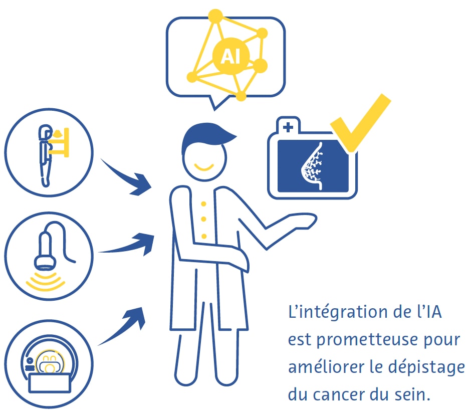
lintelligence artificielle

Références 

Aggarwal, R., Sounderajah, V., Martin, G., Ting, D. S. W., Karthikesalingam, A., King, D., Ashrafian, H., & Darzi, A. (2021). Diagnostic accuracy of deep learning in medical imaging: a systematic review and meta-analysis. NPJ Digital Medicine, 4(1), 65.

Alabousi, M., Zha, N., Salameh, J.-P., Samoilov, L., Sharifabadi, A. D., Pozdnyakov, A., Sadeghirad, B., Freitas, V., McInnes, M. D. F., & Alabousi, A. (2020). Digital breast tomosynthesis for breast cancer detection: a diagnostic test accuracy systematic review and meta-analysis. European Radiology, 30(4), 2058–2071.

Alkabban FM and Ferguson T. (2022). Breast Cancer, 2022. In: StatPearls [Internet]. Treasure Island (FL): StatPearls Publishing.

Alsheik, N. H., Dabbous, F., Pohlman, S. K., Troeger, K. M., Gliklich, R. E., Donadio, G. M., Su, Z., Menon, V., & Conant, E. F. (2019). Comparison of Resource Utilization and Clinical Outcomes Following Screening with Digital Breast Tomosynthesis Versus Digital Mammography: Findings From a Learning Health System. Academic Radiology, 26(5), 597–605.

Arefan, D., Mohamed, A. A., Berg, W. A., Zuley, M. L., Sumkin, J. H., & Wu, S. (2020). Deep learning modeling using normal mammograms for predicting breast cancer risk. Medical Physics, 47(1), 110–118.

Balleyguier, C., Arfi-Rouche, J., Levy, L., Toubiana, P. R., Cohen-Scali, F., Toledano, A. Y., & Boyer, B. (2017). Improving digital breast tomosynthesis reading time: A pilot multi-reader, multi-case study using concurrent Computer-Aided Detection (CAD). European Journal of Radiology, 97, 83–89.

Banks, E., Reeves, G., Beral, V., Bull, D., Crossley, B., Simmonds, M., Hilton, E., Bailey, S., Barrett, N., Briers, P., English, R., Jackson, A., Kutt, E., Lavelle, J., Rockall, L., Wallis, M. G., Wilson, M., & Patnick, J. (2006). Hormone replacement therapy and false positive recall in the Million Women Study: patterns of use, hormonal constituents and consistency of effect. Breast Cancer Research: BCR, 8(1), R8.

Batchu, S., Liu, F., Amireh, A., Waller, J., & Umair, M. (2021). A Review of Applications of Machine Learning in Mammography and Future Challenges. Oncology, 99(8), 483–490. https://doi.org/10.1159/000515698

Bazira, P. J., Ellis, H., & Mahadevan, V. (2022). Anatomy and physiology of the breast. Surgery, 40(2), 79–83. https://doi.org/10.1016/j.mpsur.2021.11.015

Berg, W. A., Campassi, C., Langenberg, P., & Sexton, M. J. (2000). Breast Imaging Reporting and Data System: interand intraobserver variability in feature analysis and final assessment. AJR. American Journal of Roentgenology, 174(6), 1769–1777.

Boyd, N. F., Guo, H., Martin, L. J., Sun, L., Stone, J., Fishell, E., Jong, R. A., Hislop, G., Chiarelli, A., Minkin, S., & Yaffe, M. J. (2007). Mammographic density and the risk and detection of breast cancer. The New England Journal of Medicine, 356(3), 227–236.

Bray, F., Ferlay, J., Soerjomataram, I., Siegel, R. L., Torre, L. A., & Jemal, A. (2018). Global cancer statistics 2018: GLOBOCAN estimates of incidence and mortality worldwide for 36 cancers in 185 countries. CA: A Cancer Journal for Clinicians, 68(6), 394–424.

Breast Cancer Statistics. (2020, October 27). Susan G. Komen®. https://www.komen.org/breast-cancer/facts-statistics/breastcancer- statistics/

Byrne, C., Schairer, C., Wolfe, J., Parekh, N., Salane, M., Brinton, L. A., Hoover, R., & Haile, R. (1995). Mammographic features and breast cancer risk: effects with time, age, and menopause status. Journal of the National Cancer Institute, 87(21), 1622–1629.

Chikarmane, S. A., Offit, L. R., & Giess, C. S. (2023). Synthetic Mammography: Benefits, Drawbacks, and Pitfalls. Radiographics: A Review Publication of the Radiological Society of North America, Inc, 43(10), e230018. https://doi.org/10.1148/rg.230018

Daniel, B. L., & Ikeda, D. M. (2011). Chapter 7 - Magnetic Resonance Imaging of Breast Cancer and MRI-Guided Breast Biopsy. In D. M. Ikeda (Ed.), Breast Imaging (Second Edition) (pp. 239–296). Mosby.

DeMartini W & Lehman C, (2008). Top Magn Reson Imaging, Jun;19(3):143-50.

Dembrower, K., Liu, Y., Azizpour, H., Eklund, M., Smith, K., Lindholm, P., & Strand, F. (2020). Comparison of a Deep Learning Risk Score and Standard Mammographic Density Score for Breast Cancer Risk Prediction. Radiology, 294(2), 265–272.

Dembrower, K., Wåhlin, E., Liu, Y., Salim, M., Smith, K., Lindholm, P., Eklund, M., & Strand, F. (2020). Effect of artificial intelligence-based triaging of breast cancer screening mammograms on cancer detection and radiologist workload: a retrospective simulation study. The Lancet. Digital Health, 2(9), e468–e474.

Dibden, A., Offman, J., Duffy, S. W., & Gabe, R. (2020). Worldwide Review and Meta-Analysis of Cohort Studies Measuring the Effect of Mammography Screening Programmes on Incidence-Based Breast Cancer Mortality. Cancers, 12(4). https://doi.org/10.3390/cancers12040976

Duffy, S. W., Morrish, O. W. E., Allgood, P. C., Black, R., Gillan, M. G. C., Willsher, P., Cooke, J., Duncan, K. A., Michell, M. J., Dobson, H. M., Maroni, R., Lim, Y. Y., Purushothaman, H. N., Suaris, T., Astley, S. M., Young, K. C., Tucker, L., & Gilbert, F. J. (2018). Mammographic density and breast cancer risk in breast screening assessment cases and women with a family history of breast cancer. European Journal of Cancer, 88, 48–56.

Food and Drug Administration. (2001). The Mammography Quality Standards Act Final Regulations: Preparing for MQSA Inspections; Final Guidance for Industry and FDA.

Freeman, K., Geppert, J., Stinton, C., Todkill, D., Johnson, S., Clarke, A., & Taylor-Phillips, S. (2021). Use of artificial intelligence for image analysis in breast cancer screening programmes: systematic review of test accuracy. BMJ, 374, n1872.

Freer, P. E. (2015). Mammographic breast density: impact on breast cancer risk and implications for screening. Radiographics: A Review Publication of the Radiological Society of North America, Inc, 35(2), 302–315.

Garrett, J. W., Li, Y., Li, K., & Chen, G.-H. (2018). Reduced anatomical clutter in digital breast tomosynthesis with statistical iterative reconstruction. Medical Physics, 45(5), 2009–2022.

Geras, K. J., Mann, R. M., & Moy, L. (2019). Artificial Intelligence for Mammography and Digital Breast Tomosynthesis: Current Concepts and Future Perspectives. Radiology, 293(2), 246–259. https://doi.org/10.1148/radiol.2019182627

Giordano, L., von Karsa, L., Tomatis, M., Majek, O., de Wolf, C., Lancucki, L., Hofvind, S., Nyström, L., Segnan, N., Ponti, A., Eunice Working Group, Van Hal, G., Martens, P., Májek, O., Danes, J., von Euler-Chelpin, M., Aasmaa, A., Anttila, A., Becker, N., … Suonio, E. (2012). Mammographic screening programmes in Europe: organization, coverage and participation. Journal of Medical Screening, 19 Suppl 1, 72–82.

Harbeck, N., Penault-Llorca, F., Cortes, J., Gnant, M., Houssami, N., Poortmans, P., Ruddy, K., Tsang, J., & Cardoso, F. (2019). Breast cancer. Nature Reviews. Disease Primers, 5(1), 66. https://doi.org/10.1038/s41572-019-0111-2

Hickman, S. E., Baxter, G. C., & Gilbert, F. J. (2021). Adoption of artificial intelligence in breast imaging: evaluation, ethical constraints and limitations. British Journal of Cancer, 125(1), 15–22.

Hinton, B., Ma, L., Mahmoudzadeh, A. P., Malkov, S., Fan, B., Greenwood, H., Joe, B., Lee, V., Kerlikowske, K., & Shepherd, J. (2019). Deep learning networks find unique mammographic differences in previous negative mammograms between interval and screen-detected cancers: a case-case study. Cancer Imaging: The Official Publication of the International Cancer Imaging Society, 19(1), 41.

Hirko, K. A., Rocque, G., Reasor, E., Taye, A., Daly, A., Cutress, R. I., Copson, E. R., Lee, D.-W., Lee, K.-H., Im, S.-A., & Park, Y. H. (2022). The impact of race and ethnicity in breast cancer-disparities and implications for precision oncology.BMC Medicine, 20(1), 72.

Hong, R., & Xu, B. (2022). Breast cancer: an up-to-date review and future perspectives. Cancer Communications, 42(10), 913–936. https://doi.org/10.1002/cac2.12358

Hubbard, R. A., Kerlikowske, K., Flowers, C. I., Yankaskas, B. C., Zhu, W., & Miglioretti, D. L. (2011). Cumulative probability of false-positive recall or biopsy recommendation after 10 years of screening mammography: a cohort study.Annals of Internal Medicine, 155(8), 481–492.

Hu, K., Ding, P., Wu, Y., Tian, W., Pan, T., & Zhang, S. (2019). Global patterns and trends in the breast cancer incidence and mortality according to sociodemographic indices: an observational study based on the global burden of diseases. BMJ Open, 9(10), e028461.

Ikeda, D. M. (Ed.). (2011a). Chapter 2 - Mammogram Interpretation. In Breast Imaging (Second Edition) (pp. 24–62). Mosby.

Ikeda, D. M. (Ed.). (2011b). Chapter 5 - Breast Ultrasound. In Breast Imaging (Second Edition) (pp. 149–193). Mosby.

James, J. J., Giannotti, E., & Chen, Y. (2018). Evaluation of a computer-aided detection (CAD)-enhanced 2D synthetic mammogram: comparison with standard synthetic 2D mammograms and conventional 2D digital mammography. Clinical Radiology, 73(10), 886–892.

Kalager, M., Zelen, M., Langmark, F., & Adami, H.-O. (2010). Effect of screening mammography on breast-cancer mortality in Norway. The New England Journal of Medicine, 363(13), 1203–1210. https://doi.org/10.1056/NEJMoa1000727

Kim, Y.-S., Park, H.-S., Lee, H.-H., Choi, Y.-W., Choi, J.-G., Kim, H. H., & Kim, H.-J. (2016). Comparison study of reconstruction algorithms for prototype digital breast tomosynthesis using various breast phantoms. La Radiologia Medica, 121(2), 81–92.

Koo, M. M., von Wagner, C., Abel, G. A., McPhail, S., Rubin, G. P., & Lyratzopoulos, G. (2017). Typical and atypical presenting symptoms of breast cancer and their associations with diagnostic intervals: Evidence from a national audit of cancer diagnosis. Cancer Epidemiology, 48, 140–146. https://doi.org/10.1016/j.canep.2017.04.010

Lång, K., Josefsson, V., Larsson, A.-M., Larsson, S., Högberg, C., Sartor, H., Hofvind, S., Andersson, I., & Rosso, A. (2023). Artificial intelligence-supported screen reading versus standard double reading in the Mammography Screening with Artificial Intelligence trial (MASAI): a clinical safety analysis of a randomised, controlled, non-inferiority, single-blinded, screening accuracy study. The Lancet Oncology, 24(8), 936–944.

Lauritzen, A. D., Rodríguez-Ruiz, A., von Euler-Chelpin, M. C., Lynge, E., Vejborg, I., Nielsen, M., Karssemeijer, N., & Lillholm, M. (2022). An Artificial Intelligence-based Mammography Screening Protocol for Breast Cancer: Outcome and Radiologist Workload.Radiology, 304(1), 41–49.

Lehman, C. D., Yala, A., Schuster, T., Dontchos, B., Bahl, M., Swanson, K., & Barzilay, R. (2019). Mammographic Breast Density Assessment Using Deep Learning: Clinical Implementation. Radiology, 290(1), 52–58.

Leibig, C., Brehmer, M., Bunk, S., Byng, D., Pinker, K., & Umutlu, L. (2022). Combining the strengths of radiologists and AI for breast cancer screening: a retrospective analysis. The Lancet. Digital Health, 4(7), e507–e519.

Lei, J., Yang, P., Zhang, L., Wang, Y., & Yang, K. (2014). Diagnostic accuracy of digital breast tomosynthesis versus digital mammography for benign and malignant lesions in breasts: a meta-analysis. European Radiology, 24(3), 595–602.

Lekadir, K., Osuala, R., Gallin, C., Lazrak, N., Kushibar, K., Tsakou, G., Aussó, S., Alberich, L. C., Marias, K., Tsiknakis, M., Colantonio, S., Papanikolaou, N., Salahuddin, Z., Woodruff, H. C., Lambin, P., & Martí-Bonmatí, L. (2021). FUTURE-AI: Guiding Principles and Consensus Recommendations for Trustworthy Artificial Intelligence in Medical Imaging. In arXiv [cs.CV].arXiv. https://arxiv.org/abs/2109.09658

Liu, J., Zarshenas, A., Qadir, A., Wei, Z., Yang, L., Fajardo, L., & Suzuki, K. (2018). Radiation dose reduction in digital breast tomosynthesis (DBT) by means of deep-learning-based supervised image processing. Medical Imaging 2018: Image Processing, 10574, 89–97.

Liu, X., Cruz Rivera, S., Moher, D., Calvert, M. J., Denniston, A. K., & SPIRIT-AI and CONSORT-AI Working Group. (2020). Reporting guidelines for clinical trial reports for interventions involving artificial intelligence: the CONSORT-AI extension. Nature Medicine, 26(9), 1364–1374.

Łukasiewicz, S., Czeczelewski, M., Forma, A., Baj, J., Sitarz, R., & Stanisławek, A. (2021). Breast Cancer-Epidemiology, Risk Factors, Classification, Prognostic Markers, and Current Treatment Strategies-An Updated Review. Cancers, 13(17). https://doi.org/10.3390/cancers13174287

Lynge, E., Vejborg, I., Andersen, Z., von Euler-Chelpin, M., & Napolitano, G. (2019). Mammographic Density and Screening Sensitivity, Breast Cancer Incidence and Associated Risk Factors in Danish Breast Cancer Screening. Journal of Clinical Medicine Research, 8(11). https://doi.org/10.3390/jcm8112021

Mann, R. M., Cho, N., & Moy, L. (2019). Breast MRI: State of the Art. Radiology, 292(3), 520–536.

Martini, R., Newman, L., & Davis, M. (2022). Breast cancer disparities in outcomes; unmasking biological determinants associated with racial and genetic diversity. Clinical & Experimental Metastasis, 39(1), 7–14.

Mascara, M., & Constantinou, C. (2021). Global Perceptions of Women on Breast Cancer and Barriers to Screening. Current Oncology Reports, 23(7), 74.

McDonald, E. S., Clark, A. S., Tchou, J., Zhang, P., & Freedman, G. M. (2016). Clinical Diagnosis and Management of Breast Cancer. Journal of Nuclear Medicine: Official Publication, Society of Nuclear Medicine, 57 Suppl 1, 9S – 16S. https://doi.org/10.2967/jnumed.115.157834

McKinney, S. M., Sieniek, M., Godbole, V., Godwin, J., Antropova, N., Ashrafian, H., Back, T., Chesus, M., Corrado, G. S., Darzi, A., Etemadi, M., Garcia-Vicente, F., Gilbert, F. J., Halling-Brown, M., Hassabis, D., Jansen, S., Karthikesalingam, A., Kelly, C. J., King, D., … Shetty, S. (2020). International evaluation of an AI system for breast cancer screening. Nature, 577(7788), 89–94.

Mohamed, A. A., Berg, W. A., Peng, H., Luo, Y., Jankowitz, R. C., & Wu, S. (2018). A deep learning method for classifying mammographic breast density categories. Medical Physics, 45(1), 314–321.

Moran, S., & Warren-Forward, H. (2012). The Australian BreastScreen workforce: a snapshot. The Radiographer, 59(1), 26–30.

Nelson, H. D., Fu, R., Cantor, A., Pappas, M., Daeges, M., & Humphrey, L. (2016). Effectiveness of Breast Cancer Screening: Systematic Review and Meta-analysis to Update the 2009 U.S. Preventive Services Task Force Recommendation. Annals of Internal Medicine, 164(4), 244–255.

Nelson, H. D., Pappas, M., Cantor, A., Griffin, J., Daeges, M., & Humphrey, L. (2016). Harms of Breast Cancer Screening: Systematic Review to Update the 2009 U.S. Preventive Services Task Force Recommendation. Annals of Internal Medicine, 164(4), 256–267.

Nguyen, H. T., Nguyen, H. Q., Pham, H. H., Lam, K., Le, L. T., Dao, M., & Vu, V. (2023). VinDr-Mammo: A large-scale benchmark dataset for computer-aided diagnosis in full-field digital mammography. Scientific Data, 10(1), 277.

Ong, M.-S., & Mandl, K. D. (2015). National expenditure for false-positive mammograms and breast cancer overdiagnoses estimated at $4 billion a year. Health Affairs , 34(4), 576–583.

Prevedello, L. M., Halabi, S. S., Shih, G., Wu, C. C., Kohli, M. D., Chokshi, F. H., Erickson, B. J., Kalpathy-Cramer, J., Andriole, K. P., & Flanders, A. E. (2019). Challenges Related to Artificial Intelligence Research in Medical Imaging and the Importance of Image Analysis Competitions. Radiology. Artificial Intelligence, 1(1), e180031. https://doi.org/10.1148/ ryai.2019180031

Raya-Povedano, J. L., Romero-Martín, S., Elías-Cabot, E., Gubern-Mérida, A., Rodríguez-Ruiz, A., & Álvarez-Benito, M. (2021). AI-based Strategies to Reduce Workload in Breast Cancer Screening with Mammography and Tomosynthesis: A Retrospective Evaluation. Radiology, 300(1), 57–65.

Rimmer, A. (2017). Radiologist shortage leaves patient care at risk, warns royal college. BMJ, 359, j4683.

Skaane, P., Bandos, A. I., Niklason, L. T., Sebuødegård, S., Østerås, B. H., Gullien, R., Gur, D., & Hofvind, S. (2019). Digital Mammography versus Digital Mammography Plus Tomosynthesis in Breast Cancer Screening: The Oslo Tomosynthesis Screening Trial. Radiology, 291(1), 23–30.

Sung H et al,(2021). CA Cancer J Clin, 2021 May;71(3):209-249. doi: 10.3322/caac.21660

Tabár, L., Dean, P. B., Chen, T. H.-H., Yen, A. M.-F., Chen, S. L.- S., Fann, J. C.-Y., Chiu, S. Y.-H., Ku, M. M.-S., Wu, W. Y.-Y., Hsu, C.-Y., Chen, Y.-C., Beckmann, K., Smith, R. A., & Duffy, S. W. (2019). The incidence of fatal breast cancer measures the increased effectiveness of therapy in women participating in mammography screening. Cancer, 125(4), 515–523.

Svahn, T. M., Houssami, N., Sechopoulos, I., & Mattsson, S. (2015). Review of radiation dose estimates in digital breast tomosynthesis relative to those in two-view full-field digital mammography. Breast, 24(2), 93–99. https://doi.org/10.1016/j. breast.2014.12.002

Tirada, N., Li, G., Dreizin, D., Robinson, L., Khorjekar, G., Dromi, S., & Ernst, T. (2019). Digital Breast Tomosynthesis: Physics, Artifacts, and Quality Control Considerations. Radiographics: A Review Publication of the Radiological Society of North America, Inc, 39(2), 413–426.

Torres-Mejía, G., De Stavola, B., Allen, D. S., Pérez-Gavilán, J. J., Ferreira, J. M., Fentiman, I. S., & Dos Santos Silva, I. (2005). Mammographic features and subsequent risk of breast cancer: a comparison of qualitative and quantitative evaluations in the Guernsey prospective studies. Cancer Epidemiology, Biomarkers & Prevention: A Publication of the American Association for Cancer Research, Cosponsored by the American Society of Preventive Oncology, 14(5), 1052–1059.

Volpara Health. (2022). TruPGMI: AI for mammography quality improvement. https://www.volparahealth.com/breast-healthsoftware/ products/analytics/

Wing, P., & Langelier, M. H. (2009). Workforce shortages in breast imaging: impact on mammography utilization. AJR. American Journal of Roentgenology, 192(2), 370–378.

Yala, A., Lehman, C., Schuster, T., Portnoi, T., & Barzilay, R. (2019). A Deep Learning Mammography-based Model for Improved Breast Cancer Risk Prediction. Radiology, 292(1), 60–66.

Yala, A., Schuster, T., Miles, R., Barzilay, R., & Lehman, C. (2019). A Deep Learning Model to Triage Screening Mammograms: A Simulation Study. Radiology, 293(1), 38–46.


Artificial Intelligence in medical imaging: What, How and Why?

    Artificial intelligence (AI) is a field that enables computer systems to solve problems by adapting to changing circumstances, often by mimicking human reasoning and judgement. Several demographic and healthcare trends are driving the use of AI in medical imaging. The amount of medical imaging data being acquired is steadily increasing (Larson et al., 2011; Smith-Bindman et al., 2008, 2012; Winder et al., 2021). There is also a widespread shortage of healthcare workers (Core Health Indicators in the WHO European Region 2015. Special Focus: Human Resources for Health, 2017) with an ever-increasing workload (Levin et al., 2017), and the number of medical imaging examinations is expected to grow exponentially over the next two decades (Tsao, 2020). Radiologists and radiology technologists are in particularly scarce supply (AAMC Report Reinforces Mounting Physician Shortage, 2021, Clinical Radiology UK Workforce Census 2019 Report, 2019). Finally, the ageing world population (Population Ages 65 and above, n.d.; WHO, n.d.-a) and an increasing global burden of chronic illnesses (WHO, n.d.-b) are expected to compound these problems in the near future.

    Broadly speaking, the advantages of AI in medical imaging could potentially include the ability to provide insights that would otherwise not be possible using traditional methods (such as humans looking at images) and to may do so in a faster and automated way (without the need for human interaction). AI-based solutions in medical imaging could improve and accelerate the detection of disease, generate in-depth risk assessment of disease development and progression, and may reduce subjectivity in the interpretation of medical imaging data.

    Over the past few years, the landscape of AI in medical imaging has changed dramatically. Many promising applications have arisen, the field has seen an unprecedented surge in funding, and we have seen positive trends in the adoption of AI solutions by radiologists, as well as their approval by regulatory bodies.

    Applications

    Although radiology departments provide a plethora of services, the core service provided is the imaging study. Applications of AI in medical imaging can therefore be categorized into those applied either before, during, or after the imaging study.

    Before Image Acquisition

    Several steps have to take place within the context of a radiology department’s workflow before a patient is undergoing imaging study. AI applications that aim to improve these steps are referred to as “upstream AI” and could potentially increase efficiency and provide more personalized decision making in a radiology department.

    Missed medical appointments are common, reduce the efficiency of hospitals, and waste resources (Dantas et al., 2018). Studies from Japan (Kurasawa et al., 2016) and the United Kingdom (Nelson et al., 2019) have shown that AI can be used to predict no-shows with high accuracy. This allows the use of targeted strategies to reduce the likelihood of a patient missing their appointment, including sending automated reminders.

    One of the most important decisions made in the radiology department is the exact scan protocol to use on a given patient. While this applies to all imaging modalities, the widest range of choice is seen with magnetic resonance imaging (MRI). This includes choosing the appropriate set of sequences and making decisions about whether or not to administer intravenous contrast agents. Natural language classifiers that interpret the narrative text of the clinician’s scan requests have been used to select appropriate MRI protocols. In one study, a gradient boosting classifier predicted the appropriate MRI brain protocol to use based on the scan request with high accuracy (95 %) (Brown & Marotta, 2018). For musculoskeletal MRI, a deep learning classifier was 83 % accurate in determining the need for a contrast agent (Trivedi et al., 2018). Such applications can substantially improve efficiency by foregoing the time-consuming task of radiologists going through unstructured narrative scan requests written by referring clinicians.

    During Image Acquisition

    Substantial improvements have recently been made in the use of AI for improving image quality. In a recent survey, radiologists identified the enhancement of image quality as being the most mainstream current use case for AI in medical imaging (Alexander et al., 2020). While earlier attempts at reducing image noise using deep learning techniques were criticized for removing details from the images that jeopardized the visibility of essential features within the images, more recent implementations have made this issue largely obsolete.


    Potentials of AI


    In particular, deep learning techniques like generative adversarial networks have shown great potential in image denoising (Wang et al., 2021). Some of these applications target the image reconstruction stage (where the raw sensor data is converted into an interpretable image) providing superior signal-to-noise ratios and reducing image artefacts (Zhu et al., 2018). In lung cancer screening, deep-learning-based image denoising improved both the image quality and the diagnostic accuracy of ultra-low-dose computed tomography (CT) for detecting suspicious lung nodules (Hata et al., 2020; Kerpel et al., 2021). Scans that were 40-60 % acquired faster than standard scans and enhanced with deep-learning-based algorithms were of better image quality than, and similar diagnostic value as, standard scans of the brain (Bash, Wang, et al., 2021; Rudie et al., 2022) and spine (Bash, Johnson, et al., 2021). Similarly, convolutional neural networks can be used to reduce specific CT and MRI artefacts and improve spatial resolution (Hauptmann et al., 2019; K. H. Kim & Park, 2017; Park et al., 2018; Y.Zhang & Yu, 2018).

    Reconstruction algorithms based on deep learning have enabled ultra-low-dose computed tomography scans to be acquired while maintaining diagnostic quality. This is of particular benefit in children and pregnant women, where reduction of radiation dose to the absolute minimum is critical. These deep- learning-based CT image reconstruction approaches are associated with lower image noise and better image texture than state-of-the-art alternatives like iterative reconstruction (Higaki et al., 2020; McLeavy et al., 2021; Singh et al., 2020). In positron emission tomography, deep learning can reduce injected tracer dosage by one-third and scan times by up to half while maintaining scan quality (Katsari et al., 2021; Le et al., 2020; Xu et al., 2020).

    After Image Acquisition

    Radiology technologists and radiologists usually share the task of calling back patients for repeat examinations, but doing so consistently and reliably is exceedingly difficult due to time constraints. Image quality of AI enhanced brain MRI scans has been shown to be equal to or better than conventional scans, even when using acquisition protocols that reduce scan times by 45-60% (Schreiber-Zinaman & Rosenkrantz, 2017).

    Prioritizing scan reading on a radiologist’s worklist is often done based on several factors including the type of scan, the referring department, and direct communication with the radiologist about the scan’s urgency. Several approaches have been tested to influence the order in which scans are read to improve efficiency and ensure the most critical scans are seen first. These include assigning different radiologists specific exams based on how quickly they read certain scan types (Wong et al., 2019) and automatically detecting emergent findings on the images and pushing these cases to the “top of the list” (Prevedello et al., 2017; Winkel et al., 2019).

    About 70 % of all AI-based solutions in radiology focus on “perception” - a category of functionalities that includes segmentation, feature extraction, as well as detection and classification of pathology (Rezazade Mehrizi et al., 2021). Within this category, the majority of tools extract information from the imaging data with or without quantification as well as draw the user’s attention to potential pathology (Rezazade Mehrizi et al., 2021; van Leeuwen et al., 2021). Over the past few years, some of the most promising applications in this category have included the detection of brain vessel occlusion, brain haemorrhage, lung nodules, pneumothorax and pleural effusions, fractures, and the characterization of breast lesions.

    Funding

    The total amount of investment in AI-based medical imaging companies amounted to $ 1.17 billion between 2014 and 2019 (Alexander et al., 2020). In the same period, the number of companies in this space tripled, leading to a drop of almost 30 % in the median investment in each company (Alexander et al., 2020). Between 2019 and 2020, private investment in AI companies increased by 9.3 % (D. Zhang et al., 2021). By 2030, investment in AI-based solutions in medical imaging is expected to exceed $3 billion (Tsao, 2020).

    Adoption

    There have been positive trends in the adoption of AI tools by radiologists and radiology technologists over the past few years. Between 2015 and 2020, AI use in radiology departments went up by 30 %, according to a survey of 1,861 radiologists conducted by the American College of Radiology (ACR) (Allen et al., 2021).

    Despite this promising trend, the adoption of AI tools is widely considered to be disproportionately low relative to the amount of funding, the number of companies, and the perceived promise of these tools. The ACR survey provides some insight into why and offers a starting point for developing strategies to improve AI adoption.

    Almost three-quarters of radiologists who were not using AI had no plans to do so in the future because they either were not convinced of its benefits or did not think the associated costs were justified (Allen et al., 2021).

    Similar results have been found in other studies, with radiologists citing scepticism in the capabilities of AI tools and the fact that relatively few have regulatory approval as reasons for not adopting them in their practice (Alexander et al., 2020).

    Regulatory success

    Until August 2019, 60 % of available AI-based radiology solutions had no regulatory approval (Rezazade Mehrizi et al., 2021). As of April 2020, a total of 100 AI solutions had a CE mark, a prerequisite for them to be made commercially available as medical devices in Europe (van Leeuwen et al., 2021). As of the time of writing, more than 150 AI solutions have gained FDA clearance (AI Central, n.d.). Several useful databases of approved or cleared AI-based solutions in medical healthcare are currently available (AI Central, n.d., AI for Radiology, n.d., Medical AI Evaluation, n.d., The Medical Futurist, n.d.)
     

    The past few years have seen exponential growth in the interest in AI in medical imaging, both in terms of the amount of research and the amount of money being invested in the field. This interest runs the gamut of the radiology workflow, but “perception” applications - for the quantification of biomarkers and the detection of disease processes - have dominated so far. In the radiology community, trends have shifted from AI being perceived as an unwelcome intruder to increased adoption, albeit with some scepticism and hesitation regarding its value. The first AI solutions in medical imaging were granted regulatory approval, and we have seen the first indications of how such solutions may be reimbursed.

    New directions

    With increasing acknowledgement that a large proportion of AI’s potential in medical imaging may lie in “upstream” or “non-interpretative” applications, the field is likely to expand its focus in the coming years. This will include more research into applications that improve the efficiency of radiology workflows and provide more personalized patient care (Alexander et al., 2020). AI is likely to become more involved even earlier in the patient management process - i.e. before the clinician decides that a diagnostic image test is necessary. Such applications, essentially clinical decision support systems, have successfully been used for decision-making about treatments in several settings (Bennett & Hauser, 2013; Komorowski et al., 2018), successfully used in treatment decision making (Bennett & Hauser, 2013). In the future, AI solutions may draw clinicians’ attention to the need for further imaging tests based on reviewing the patient’s clinical information, laboratory tests, and prior imaging tests (Makeeva et al., 2019).


    Perception of AI

    The vast majority (77-84 %) of currently available AI solutions in medical imaging target CT, MRI and plain radiographs (Rezazade Mehrizi et al., 2021; van Leeuwen et al., 2021). Nuclear imaging techniques, such as positron emission tomography (PET). provide unique information not readily gained from other modalities. PET has thus far been largely neglected in terms of AI research (Rezazade Mehrizi et al., 2021; van Leeuwen et al., 2021), and is thus a potentially promising avenue for the field’s expansion.

    AI research is also expected to undergo a shift in the type of data being used. The typical inpatient receives more than one imaging study during their hospital stay (Shinagare et al., 2014). Despite this, only about 3 % of current AI-based radiology solutions combine data from multiple modalities (Rezazade Mehrizi et al., 2021; van Leeuwen et al., 2021). Combining data from multiple imaging sources may improve the diagnostic capabilities of AI solutions. Furthermore, future AI solutions in radiology are likely to combine imaging information, clinical information, as well as non- imaging diagnostic tests (Huang et al., 2020). By doing this, AI solutions may be able to identify patterns in the data collected during a patient’s hospital stay that may not be readily identifiable by healthcare workers (Rockenbach, 2021). This could ultimately lead to more accurate diagnoses and could help inform better and more personalized treatment decisions.

    The expectations for AI-based medical imaging solutions are also likely to shift from the current focus of triage, image enhancement and automation. With increasing algorithmic complexity, data availability, and experience with these tools, this shift may lead to AI solutions reaching specific diagnoses and recommending specific steps in a patient’s management plan. Similar to how the introduction of the first AI tools for image screening and processing around 2018 spurred investment in the field, marketing analyses predict a similar investment boost in the next few years as AI tools providing specific diagnoses and management steps become more widespread (Michoud et al., 2019).

    One important criticism of the current, arguably still nascent, landscape of AI in medical imaging is that it is too fragmented. Radiology professionals would likely welcome a more streamlined integration of AI solutions in their daily workflow. This includes seamless integration of these solutions into established radiology workflows, with as much as possible happening “in the background” without user input. Furthermore, the outputs of these solutions could be integrated into available radiological information systems. Consequently, the field could move from the plethora of currently available niche AI solutions, each targeted towards a single very specific application, to broader software suites that perform many different functions for a given imaging modality or body region.

    The fragmented investment in the AI in medical imaging market (Alexander et al., 2020) fosters innovation, allowing many players to test out different strategies in this emerging field. However, in the long term, consolidation may increase adoption and stimulate the kind of seamless integration into existing workflows that is needed, allowing fewer companies to offer these solutions at scale (Alexander et al., 2020).

    Challenges

    Quality and reporting of evidence

    In a review of 100 CE-marked AI solutions, 64 % of them had no peer-reviewed scientific evidence for their efficacy (van Leeuwen et al., 2021). Where there was scientific evidence, the level was low, rarely exceeding the demonstration of diagnostic accuracy (van Leeuwen et al., 2021). Another systematic review of the evidence for deep learning algorithms in medical imaging found a generally high diagnostic accuracy, albeit with a high risk of bias across studies (Aggarwal et al., 2021). The main sources of bias include the lack of external validation (D. W. Kim et al., 2019; Liu et al., 2019), insufficiently detailed reporting of results (Liu et al., 2019), retrospective study design (Nagendran et al., 2020), and the inaccessibility of data and code to reviewers and readers (Nagendran et al., 2020).

    Overall, studies on AI tools have shown a worrying lack of standardized reporting and adherence to recommended reporting guidelines (Aggarwal et al., 2021; Yusuf et al., 2020). This is despite the fact that several extensions to established reporting guidelines, as well as AI-specific guidelines, are currently available (Shelmerdine et al., 2021). Widespread implementation of these guidelines should be a focus of AI developers in the future.

    AI developers should also be cognizant that the currently “acceptable” level of evidence for AI-based solutions is likely to become obsolete in the near future. Both regulators and potential users will likely demand higher levels of evidence for these solutions, akin to the evidence required for new pharmaceutical drugs. In the next few years, will see more of these AI solutions being tested in randomized clinical trials. In the more distant future, it is plausible that such expectations will go beyond providing evidence of the safety, efficacy, or diagnostic performance of these solutions, to the demonstration that they provide added monetary or societal value.

    Rising up to the challenge of improving the quality and reporting of evidence for AI-based solutions may pay off in the long run. It could reduce the risk of bias in AI studies, could allow the thorough and transparent assessment of study quality by potential users and regulators, and could facilitate systematic reviews and meta-analyses. These steps may increase the trust in, and uptake of, AI-based solutions and ensure that they offer realistic, sustainable improvements in people’s lives.

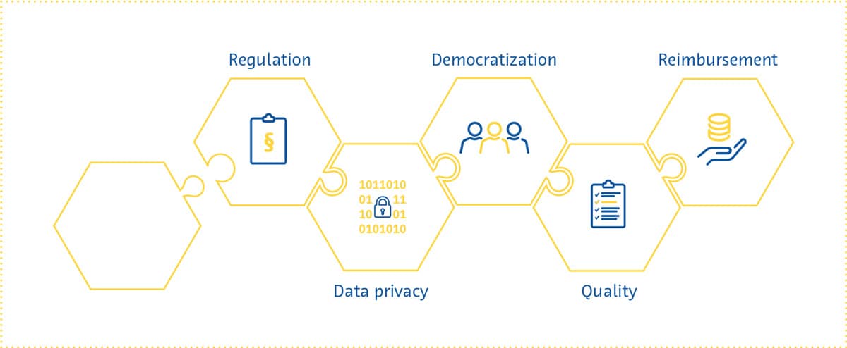
    Regulation

    Several aspects inherent to AI pose challenges to attempts at regulating it like other healthcare interventions. The inner workings of AI solutions are often opaque and difficult to comprehensively describe in a manner traditionally expected by regulatory bodies.

    The past few years have shown us that these regulatory challenges are far from intractable. Both the Food and Drug Administration and the European Commission have recently proposed initial regulatory frameworks for AI solutions (Center for Devices & Radiological Health, 2021; European Commission, 2021).

    In part as a response to the transparency necessary for regulatory approval, researchers have made substantial progress in making AI’s decision-making more understandable and explainable. This movement towards “interpretable AI” will gain further impetus in the near future as reliance on AI for real-world clinical decision-making increases.

    This has many advantages, including making regulatory approval easier, increasing trust in these solutions by users, minimizing biases, and improving the reproducibility of these solutions (Holzinger et al., 2017; Kolyshkina & Simoff, 2021; “Towards Trustable Machine Learning,” 2018; Yoon et al., 2021).

    Data privacy

    From development and testing to implementation, AI solutions in medical imaging require access to patient data. This has raised concerns about data privacy, which is a multifaceted and highly complex issue (Murdoch, 2021) that is prominently represented in the regulatory pathways of different countries (COCIR, the European Coordination Committee of the Radiological, Electromedical and Healthcare IT Industry, 2020). Suggested solutions to the data privacy question have ranged from those focusing on oversight to more technical approaches.

    The patients providing the data have to be made aware that they are doing so, as well as be informed about why and how their data will be used (Lotan et al., 2020), as explicitly stipulated in the EU’s General Data Protection Regulation (GDPR) (General Data Protection Regulation (GDPR) – Official Legal Text, 2016). Considering the fast-paced nature of the development of AI solutions, whether patients can be kept sufficiently informed as these algorithms are continuously retrained has been questioned (Kritikos, 2020). While fully anonymized data is not subject to such strict requirements under the GDPR (What Is Personal Data?, 2021), anonymization is exceedingly difficult to achieve for medical imaging data.

    The data privacy issue will have to be approached on several fronts. In addition to legislation governing the use of patient data, it is becoming increasingly clear that everyone involved in the development and use of AI solutions - developers, payers, regulatory bodies, researchers and radiologists - has a role to play in ensuring that the data is protected and used responsibly.

    Moreover, the next few years will likely see further research into technical approaches to strengthen data protection. These include better ways to reduce the chances of data being traced back to individuals, methods for keeping sensitive data stored locally even when the algorithm being trained is hosted in some “central” location, data perturbation to minimize the information within a given dataset pertaining to individual patients, and data encryption (G. Kaissis et al., 2021; G. A. Kaissis et al., 2020).

    hexagon


    Democratization

    If AI in medical imaging is to live up to its potential, the algorithms being developed have to work for everyone. This “democratization” of AI involves ensuring that healthcare providers have the knowledge and skills needed to use AI-based solutions. With a few exceptions (Paranjape et al., 2019), medical student curricula currently include little to no dedicated education about AI (Banerjee et al., 2021; Blease et al., 2022). Surveys from around the world have shown that medical students’ and doctors’ (Ahmed et al., 2022; Bisdas et al., 2021; Collado-Mesa et al., 2018; Kansal et al., 2022; Pinto Dos Santos et al., 2019; Sit et al., 2020) exposure to AI during training was low despite the high demand for more AI education (Kansal et al., 2022; Ooi et al., 2021; Sit et al., 2020). In addition, there are still large differences between genders and countries in the perceived knowledge about AI amongst medical students (Bisdas et al., 2021). There are many reasons for these differences and many challenges associated with the widespread integration of AI education into healthcare training curricula. In the coming years, strategies to tackle these issues should be investigated to ensure that future healthcare providers are equipped with the knowledge and skills they need to work in an environment where AI plays a growing role.

    Democratization also involves ensuring that patients of different genders, lifestyles, ethnicities, and geographical locations can benefit from AI-based solutions. For this to happen, these solutions have to be accessible and their performance generalizable. The latter requires the acquisition of diverse data from multiple institutions, preferably from multiple countries, for training AI-based solutions. It also requires the implementation of safeguards to ensure that sources of bias throughout the development process are not propagated to the trained algorithm (Vokinger et al., 2021), an issue that has only recently come to the forefront (Larrazabal et al., 2020; Obermeyer et al., 2019; Seyyed-Kalantari et al., 2021).

    Reimbursement

    As countries’ policies for regulating AI in healthcare gradually begin to take shape, one important aspect that needs attention is who will pay for these AI solutions, and according to what framework.

    Many consider Germany’s 2020 Digital Supply Act a step in the right direction for reimbursement of digital health solutions. Under this policy, digital applications prescribed by physicians are reimbursable by statutory health insurance if they are proven to be safe, be compliant with data privacy statutes, and improve patient care. The UK, on the other hand, has released a guide for potential buyers of AI-based solutions, which serves as a starting point for companies to prepare for reimbursement applications (A Buyer’s Guide to AI in Health and Care, 2020).

    Thus far, reimbursement success stories in the digital health space have been few and far between (Brink- mann-Sass et al., 2020; Hassan, 2021). This is in part due to requirements varying greatly by country (COCIR, the European Coordination Committee of the Radiological, Electromedical and Healthcare IT Industry, 2020). In general, providers of digital health solutions will need to provide evidence for the overall value that these solutions bring, including detailed health economics studies showing potential cost savings.

    Radiology’s position as a service provider to multiple hospital departments means that AI-based solutions in this space will be expected to show a far-reaching impact (van Duffelen, 2021). Companies will need to show short-term value (e.g. faster/better image reading and reporting) as well as long-term value (e.g. early diagnosis and treatment, disease prevention, reduction in unnecessary follow-up). The coming years will see companies compete to demonstrate such impact, while at the same time experimenting with different pricing models and navigating the evolving bureaucratic reimbursement landscape.

    Over the past few years, the field of AI in medical imaging has undergone a rapid but steady transformation. AI can now achieve things in radiology that few people thought possible a mere decade ago. The field is also gradually overcoming one of its most significant perceived hurdles - regulatory approval. In addition, while fear and scepticism dominated radiologists’ perception of the future of AI in their speciality a few years ago, this is no longer the case.

    The massive progress and interest in the field of AI in medical imaging is expected to continue into 2022 and beyond. Several exciting transformations await the field - it will likely expand its focus in the coming years to improve radiology workflow efficiency, involve hitherto neglected imaging modalities, combine data from multiple modalities, and provide more concrete diagnostic predictions and management recommendations. Easy-to-use and comprehensive software suites utilizing AI will be incorporated into existing radiology workflows, making radiologists’ and radiographers’ work easier and more efficient.

    As in any rapidly growing field, several scientific, regulatory, and economic challenges face AI in medical imaging. But the past few years have shown us that even the most difficult problems can be solved. Developers and users of AI-based solutions need to be aware of these issues so that they can adapt their strategies to changing expectations on a regulatory and societal level. Doing this will allow them to thrive in a fascinating field with the potential to improve virtually every aspect of healthcare.

    AAMC Report Reinforces Mounting Physician Shortage. (2021). AAMC. https://www.aamc.org/news-insights/press- releases/aamc-report-reinforces-mounting-physician-shortage

    A buyer’s guide to AI in health and care. (2020). NHS Transformation Directorate. https://www.nhsx.nhs.uk/ai-lab/ explore-all-resources/adopt-ai/a-buyers-guide-to-ai-in-health- and-care/

    Aggarwal, R., Sounderajah, V., Martin, G., Ting, D. S. W., Karthikesalingam, A., King, D., Ashrafian, H., & Darzi, A. (2021). Diagnostic accuracy of deep learning in medical imaging: a systematic review and meta-analysis. NPJ Digital Medicine, 4(1), 65.

    Ahmed, Z., Bhinder, K. K., Tariq, A., Tahir, M. J., Mehmood, Q., Tabassum, M. S., Malik, M., Aslam, S., Asghar, M. S., & Yousaf, Z. (2022). Knowledge, attitude, and practice of artificial intelligence among doctors and medical students in Pakistan: A cross-sectional online survey. Annals of Medicine and Surgery (2012), 76, 103493.

    AI Central. (n.d.). Retrieved February 23, 2022, from https://aicentral.acrdsi.org/

    AI for Radiology. (n.d.). Retrieved February 23, 2022, from https://grand-challenge.org/aiforradiology/

    Alexander, A., Jiang, A., Ferreira, C., & Zurkiya, D. (2020). An Intelligent Future for Medical Imaging: A Market Outlook on Artificial Intelligence for Medical Imaging. Journal of the American College of Radiology: JACR, 17(1 Pt B), 165–170.

    Allen, B., Agarwal, S., Coombs, L., Wald, C., & Dreyer, K. (2021). 2020 ACR Data Science Institute Artificial Intelligence Survey. Journal of the American College of Radiology: JACR, 18(8), 1153–1159.

    Banerjee, M., Chiew, D., Patel, K. T., Johns, I., Chappell, D., Linton, N., Cole, G. D., Francis, D. P., Szram, J., Ross, J., & Zaman, S. (2021). The impact of artificial intelligence on clinical education: perceptions of postgraduate trainee doctors in London (UK) and recommendations for trainers. BMC Medical Education, 21(1), 429.

    Bash, S., Johnson, B., Gibbs, W., Zhang, T., Shankaranarayanan, A., & Tanenbaum, L. N. (2021). Deep Learning Image Processing Enables 40 % Faster Spinal MR Scans Which Match or Exceed Quality of Standard of Care : A Prospective Multicenter Multireader Study. Clinical Neuroradiology. https://doi.org/10.1007/s00062-021-01121-2

    Bash, S., Wang, L., Airriess, C., Zaharchuk, G., Gong, E., Shankaranarayanan, A., & Tanenbaum, L. N. (2021). Deep Learning Enables 60 % Accelerated Volumetric Brain MRI While Preserving Quantitative Performance: A Prospective, Multicenter, Multireader Trial. AJNR. American Journal of Neuroradiology, 42(12), 2130–2137.

    Bennett, C. C., & Hauser, K. (2013). Artificial intelligence framework for simulating clinical decision-making: a Markov decision process approach. Artificial Intelligence in Medicine, 57(1), 9–19.

    Bisdas, S., Topriceanu, C.-C., Zakrzewska, Z., Irimia, A.-V., Shakallis, L., Subhash, J., Casapu, M.-M., Leon-Rojas, J., Pinto Dos Santos, D., Andrews, D. M., Zeicu, C., Bouhuwaish, A. M., Lestari, A. N., Abu-Ismail, L. ’i, Sadiq, A. S., Khamees, A. ’atasim, Mohammed, K. M. G., Williams, E., Omran, A. I.,… Ebrahim, E. H. (2021). Artificial Intelligence in Medicine: A Multinational Multi-Center Survey on the Medical and Dental Students’ Perception. Frontiers in Public Health, 9, 795284.

    Blease, C., Kharko, A., Bernstein, M., Bradley, C., Houston, M., Walsh, I., Hägglund, M., DesRoches, C., & Mandl, K. D. (2022). Machine learning in medical education: a survey of the experiences and opinions of medical students in Ireland. BMJ Health & Care Informatics, 29(1). https://doi.org/10.1136/ bmjhci-2021-100480

    Brown, A. D., & Marotta, T. R. (2018). Using machine learning for sequence-level automated MRI protocol selection in neuroradiology. Journal of the American Medical Informatics Association: JAMIA, 25(5), 568–571.

    Center for Devices, & Radiological Health. (2021, June 22). Digital Health Software Precertification (Pre-Cert) program.
    U.S. Food and Drug Administration. https://www.fda.gov/ medical-devices/digital-health-center-excellence/digital-health- software-precertification-pre-cert-program

    Clinical radiology UK workforce census 2019 report. (2019). https://www.rcr.ac.uk/publication/clinical-radiology-uk- workforce-census-2019-report

    COCIR, the European Coordination Committee of the Radiological, Electromedical and Healthcare IT Industry. (2020). Market Access Pathways for Digital Health Solutions. https://www.cocir.org/fileadmin/Publications_2020/20062_ COCIR_Market_Access_Pathways_Digital_Health.pdf

    Collado-Mesa, F., Alvarez, E., & Arheart, K. (2018). The Role of Artificial Intelligence in Diagnostic Radiology: A Survey at a Single Radiology Residency Training Program. Journal of the American College of Radiology: JACR, 15(12), 1753–1757.

    Core Health Indicators in the WHO European Region 2015. Special focus: Human resources for health. (2017, August 14). World Health Organization. https://www.euro.who.int/en/ data-and-evidence/evidence-resources/core-health-indicators- in-the-who-european-region/core-health-indicators-in-the- who-european-region-2015.-special-focus-human-resources- for-health

    Dantas, L. F., Fleck, J. L., Cyrino Oliveira, F. L., & Hamacher, S. (2018). No-shows in appointment scheduling - a systematic literature review. Health Policy, 122(4), 412–421.

    Esses, S. J., Lu, X., Zhao, T., Shanbhogue, K., Dane, B., Bruno, M., & Chandarana, H. (2018). Automated image quality evaluation of T2 -weighted liver MRI utilizing deep learning architecture. Journal of Magnetic Resonance Imaging: JMRI, 47(3), 723–728.

    European Commission. (2021). Proposal for a Regulation Of The European Parliament And Of The Council Laying Down
    Harmonised Rules On Artificial Intelligence (Artificial Intelligence Act) And Amending Certain Union Legislative Act.
    https://eur-lex. europa.eu/resource.html?uri=cellar:e0649735-a372-11eb- 9585-01aa75ed71a1.0001.02/DOC_1&format=PDF

    General Data Protection Regulation (GDPR) – Official Legal Text. (2016, July 13). General Data Protection Regulation (GDPR). https://gdpr-info.eu/

    Hata, A., Yanagawa, M., Yoshida, Y., Miyata, T., Tsubamoto, M., Honda, O., & Tomiyama, N. (2020). Combination of Deep Learning-Based Denoising and Iterative Reconstruction for Ultra-Low-Dose CT of the Chest: Image Quality and Lung-RADS Evaluation. AJR. American Journal of Roentgenology, 215(6), 1321–1328.

    Hauptmann, A., Arridge, S., Lucka, F., Muthurangu, V., & Steeden, J. A. (2019). Real-time cardiovascular MR with
    spatio-temporal artifact suppression using deep learning-proof of concept in congenital heart disease. Magnetic Resonance in Medicine: Official Journal of the Society of Magnetic Resonance in Medicine / Society of Magnetic Resonance in Medicine, 81(2), 1143–1156.

    Higaki, T., Nakamura, Y., Zhou, J., Yu, Z., Nemoto, T., Tatsugami, F., & Awai, K. (2020). Deep Learning Reconstruction at CT: Phantom Study of the Image Characteristics. Academic Radiology, 27(1), 82–87.

    Holzinger, A., Biemann, C., Pattichis, C. S., & Kell, D. B. (2017). What do we need to build explainable AI systems for the medical domain? In arXiv [cs.AI]. arXiv. http://arxiv.org/ abs/1712.09923

    Hötker, A. M., Da Mutten, R., Tiessen, A., Konukoglu, E., & Donati, O. F. (2021). Improving workflow in prostate MRI: AI- based decision-making on biparametric or multiparametric MRI. Insights into Imaging, 12(1), 112.

    Huang, S.-C., Pareek, A., Seyyedi, S., Banerjee, I., & Lungren, M. P. (2020). Fusion of medical imaging and electronic health records using deep learning: a systematic review and implementation guidelines. NPJ Digital Medicine, 3, 136.

    Kaissis, G. A., Makowski, M. R., Rückert, D., & Braren, R. F. (2020). Secure, privacy-preserving and federated machine learning in medical imaging. Nature Machine Intelligence, 2(6), 305–311.

    Kaissis, G., Ziller, A., Passerat-Palmbach, J., Ryffel, T., Usynin, D., Trask, A., Lima, I., Mancuso, J., Jungmann, F., Steinborn, M.-M., Saleh, A., Makowski, M., Rueckert, D., & Braren, R. (2021). End-to-end privacy preserving deep learning on multi-institutional medical imaging. Nature Machine Intelligence, 3(6), 473–484.

    Kansal, R., Bawa, A., Bansal, A., Trehan, S., Goyal, K., Goyal, N., & Malhotra, K. (2022). Differences in Knowledge and Perspectives on the Usage of Artificial Intelligence Among Doctors and Medical Students of a Developing Country: A Cross- Sectional Study. Cureus, 14(1), e21434.

    Katsari, K., Penna, D., Arena, V., Polverari, G., Ianniello, A., Italiano, D., Milani, R., Roncacci, A., Illing, R. O., & Pelosi, E. (2021). Artificial intelligence for reduced dose 18F-FDG PET examinations: a real-world deployment through a standardized framework and business case assessment. EJNMMI Physics, 8(1), 25.

    Kerpel, A., Marom, E. M., Green, M., Eifer, M., Konen, E., Mayer, A., & Betancourt Cuellar, S. L. (2021). Ultra-Low Dose Chest CT with Denoising for Lung Nodule Detection. The Israel Medical Association Journal: IMAJ, 23(9), 550–555.

    Kim, D. W., Jang, H. Y., Kim, K. W., Shin, Y., & Park, S. H. (2019).
    Design Characteristics of Studies Reporting the Performance of Artificial Intelligence Algorithms for Diagnostic Analysis of
    Medical Images: Results from Recently Published Papers. Korean Journal of Radiology: Official Journal of the Korean Radiological Society, 20(3), 405–410.

    Kim, K. H., & Park, S.-H. (2017). Artificial neural network for suppression of banding artifacts in balanced steady-state free precession MRI. Magnetic Resonance Imaging, 37, 139–146.

    Kolyshkina, I., & Simoff, S. (2021). Interpretability of Machine Learning Solutions in Public Healthcare: The CRISP-ML Approach. Frontiers in Big Data, 4, 660206.

    Komorowski, M., Celi, L. A., Badawi, O., Gordon, A. C., & Faisal, A. A. (2018). The Artificial Intelligence Clinician learns optimal treatment strategies for sepsis in intensive care. Nature Medicine, 24(11), 1716–1720.

    Kritikos, M. (2020). What if artificial intelligence in medical imaging could accelerate Covid-19 treatment? Think Tank
    - European Parliament. https://www.europarl.europa.eu/ thinktank/en/document/EPRS_ATA(2020)656333

    Kurasawa, H., Hayashi, K., Fujino, A., Takasugi, K., Haga, T., Waki, K., Noguchi, T., & Ohe, K. (2016). Machine-Learning- Based Prediction of a Missed Scheduled Clinical Appointment by Patients With Diabetes. Journal of Diabetes Science and Technology, 10(3), 730–736.

    Larrazabal, A. J., Nieto, N., Peterson, V., Milone, D. H., & Ferrante, E. (2020). Gender imbalance in medical imaging datasets produces biased classifiers for computer-aided diagnosis. Proceedings of the National Academy of Sciences of the United States of America, 117(23), 12592–12594.

    Larson, D. B., Johnson, L. W., Schnell, B. M., Salisbury, S. R., & Forman, H. P. (2011). National trends in CT use in the emergency department: 1995-2007. Radiology, 258(1), 164–173.

    Le, V., Frye, S., Botkin, C., Christopher, K., Gulaka, P., Sterkel, B., Frye, R., Muzaffar, R., & Osman, M. (2020). Effect of PET Scan with Count Reduction Using AI-Based Processing Techniques on Image Quality. Journal of Nuclear Medicine: Official Publication, Society of Nuclear Medicine, 61(supplement 1), 3095–3095.

    Levin, D. C., Parker, L., & Rao, V. M. (2017). Recent Trends in Imaging Use in Hospital Settings: Implications for Future Planning. Journal of the American College of Radiology: JACR, 14(3), 331–336.

    Liu, X., Faes, L., Kale, A. U., Wagner, S. K., Fu, D. J., Bruynseels, A., Mahendiran, T., Moraes, G., Shamdas, M., Kern, C., Ledsam, J. R., Schmid, M. K., Balaskas, K., Topol, E.J., Bachmann, L. M., Keane, P. A., & Denniston, A. K. (2019). A comparison of deep learning performance against health-care professionals in detecting diseases from medical imaging: a systematic review and meta-analysis. The Lancet. Digital Health, 1(6), e271–e297.

    Lotan, E., Tschider, C., Sodickson, D. K., Caplan, A. L., Bruno, M., Zhang, B., & Lui, Y. W. (2020). Medical Imaging and Privacy in the Era of Artificial Intelligence: Myth, Fallacy, and the Future. Journal of the American College of Radiology: JACR, 17(9), 1159–1162.

    Mairhöfer, D., Laufer, M., Simon, P. M., Sieren, M., Bischof, A., Käster, T., Barth, E., Barkhausen, J., & Martinetz, T. (2021). An AI-based Framework for Diagnostic Quality Assessment of Ankle Radiographs. https://openreview.net/pdf?id=bj04hJss_xZ

    Makeeva, V., Gichoya, J., Hawkins, C. M., Towbin, A. J., Heilbrun, M., & Prater, A. (2019). The Application of Machine Learning to Quality Improvement Through the Lens of the Radiology Value Network. Journal of the American College of Radiology: JACR, 16(9 Pt B), 1254–1258.

    McLeavy, C. M., Chunara, M. H., Gravell, R. J., Rauf, A., Cushnie, A., Staley Talbot, C., & Hawkins, R. M. (2021). The future of CT: deep learning reconstruction. Clinical Radiology, 76(6), 407–415.

    Medical AI Evaluation. (n.d.). Retrieved February 23, 2022, from https://ericwu09.github.io/medical-ai-evaluation/

    Michoud, L., Tschudi, Y., & Villien, Y. (2019). Artificial Intelligence for Medical Imaging: Market and Technology Report 2020. Yole Développement. https://s3.i-micronews.com/ uploads/2020/01/YDR20059-AI-for-Medical-Imaging_Yole_ sample.pdf

    Murdoch, B. (2021). Privacy and artificial intelligence: challenges for protecting health information in a new era. BMC Medical Ethics, 22(1), 122.

    Nagendran, M., Chen, Y., Lovejoy, C. A., Gordon, A. C., Komorowski, M., Harvey, H., Topol, E. J., Ioannidis, J. P. A., Collins, G. S., & Maruthappu, M. (2020). Artificial intelligence versus clinicians: systematic review of design, reporting standards, and claims of deep learning studies. BMJ, 368. https://doi.org/10.1136/bmj.m689

    Nelson, A., Herron, D., Rees, G., & Nachev, P. (2019). Predicting scheduled hospital attendance with artificial intelligence. Npj Digital Medicine, 2(1), 26.

    Obermeyer, Z., Powers, B., Vogeli, C., & Mullainathan, S. (2019). Dissecting racial bias in an algorithm used to manage the health of populations. Science, 366(6464), 447–453.

    Ooi, S. K. G., Makmur, A., Soon, A. Y. Q., Fook-Chong, S., Liew, C., Sia, S. Y., Ting, Y. H., & Lim, C. Y. (2021). Attitudes toward artificial intelligence in radiology with learner needs assessment within radiology residency programmes: a national multi-programme survey. Singapore Medical Journal, 62(3), 126–134.

    Paranjape, K., Schinkel, M., Nannan Panday, R., Car, J., & Nanayakkara, P. (2019). Introducing Artificial Intelligence Training in Medical Education. JMIR Medical Education, 5(2), e16048.

    Park, J., Hwang, D., Kim, K. Y., Kang, S. K., Kim, Y. K., & Lee, J. S. (2018). Computed tomography super-resolution using deep convolutional neural network. Physics in Medicine and Biology, 63(14), 145011.

    Pinto Dos Santos, D., Giese, D., Brodehl, S., Chon, S. H., Staab, W., Kleinert, R., Maintz, D., & Baeßler, B. (2019). Medical students’ attitude towards artificial intelligence: a multicentre survey. European Radiology, 29(4), 1640–1646.

    Population ages 65 and above. (n.d.). The World Bank. Retrieved February 23, 2022, from https://data.worldbank.org/ indicator/SP.POP.65UP.TO.ZS

    Prevedello, L. M., Erdal, B. S., Ryu, J. L., Little, K. J., Demirer, M., Qian, S., & White, R. D. (2017). Automated Critical Test Findings Identification and Online Notification System Using Artificial Intelligence in Imaging. Radiology, 285(3), 923–931.

    Rezazade Mehrizi, M. H., van Ooijen, P., & Homan, M. (2021). Applications of artificial intelligence (AI) in diagnostic radiology: a technography study. European Radiology, 31(4), 1805–1811.

    Rockenbach, M. A. B. (2021, June 13). Multimodal AI in healthcare: Closing the gaps. CodeX. https://medium.com/codex/ multimodal-ai-in-healthcare-1f5152e83be2

    Rudie, J. D., Gleason, T., Barkovich, M. J., Wilson, D. M., Shankaranarayanan, A., Zhang, T., Wang, L., Gong, E., Zaharchuk, G., & Villanueva-Meyer, J. E. (2022). Clinical Assessment of Deep Learning–based Super-Resolution for 3D Volumetric Brain MRI. Radiology: Artificial Intelligence, e210059.

    Schreiber-Zinaman, J., & Rosenkrantz, A. B. (2017). Frequency and reasons for extra sequences in clinical abdominal MRI examinations. Abdominal Radiology (New York), 42(1), 306–311.

    Seyyed-Kalantari, L., Zhang, H., McDermott, M. B. A., Chen, I. Y., & Ghassemi, M. (2021). Underdiagnosis bias of artificial intelligence algorithms applied to chest radiographs in under-served patient populations. Nature Medicine, 27(12), 2176–2182.

    Shelmerdine, S. C., Arthurs, O. J., Denniston, A., & Sebire, N. J. (2021). Review of study reporting guidelines for clinical studies using artificial intelligence in healthcare. BMJ Health & Care Informatics, 28(1). https://doi.org/10.1136/ bmjhci-2021-100385

    Shinagare, A. B., Ip, I. K., Abbett, S. K., Hanson, R., Seltzer, S. E., & Khorasani, R. (2014). Inpatient imaging utilization: trends of the past decade. AJR. American Journal of Roentgenology, 202(3), W277–W283.

    Singh, R., Digumarthy, S. R., Muse, V. V., Kambadakone, A. R., Blake, M. A., Tabari, A., Hoi, Y., Akino, N., Angel, E., Madan, R., & Kalra, M. K. (2020). Image Quality and Lesion Detection on Deep Learning Reconstruction and Iterative Reconstruction of Submillisievert Chest and Abdominal CT. AJR. American Journal of Roentgenology, 214(3), 566–573.

    Sit, C., Srinivasan, R., Amlani, A., Muthuswamy, K., Azam, A., Monzon, L., & Poon, D. S. (2020). Attitudes and perceptions of UK medical students towards artificial intelligence and radiology: a multicentre survey. Insights into Imaging, 11(1), 14.

    Smith-Bindman, R., Miglioretti, D. L., Johnson, E., Lee, C., Feigelson, H. S., Flynn, M., Greenlee, R. T., Kruger, R. L., Hornbrook, M. C., Roblin, D., Solberg, L. I., Vanneman, N., Weinmann, S., & Williams, A. E. (2012). Use of diagnostic imaging studies and associated radiation exposure for patients enrolled in large integrated health care systems, 1996-2010. JAMA: The Journal of the American Medical Association, 307(22), 2400–2409.

    Smith-Bindman, R., Miglioretti, D. L., & Larson, E. B. (2008). Rising use of diagnostic medical imaging in a large integrated health system. Health Affairs, 27(6), 1491–1502.

    The Medical Futurist. (n.d.). The Medical Futurist. Retrieved February 23, 2022, from https://medicalfuturist.com/fda- approved-ai-based-algorithms/

    Towards trustable machine learning. (2018). Nature Biomedical Engineering, 2(10), 709–710.

    Trivedi, H., Mesterhazy, J., Laguna, B., Vu, T., & Sohn, J. H. (2018). Automatic Determination of the Need for Intravenous Contrast in Musculoskeletal MRI Examinations Using IBM Watson’s Natural Language Processing Algorithm. Journal of Digital Imaging, 31(2), 245–251.

    Tsao, D. N. (2020, July 27). AI in medical diagnostics 2020- 2030: Image recognition, players, clinical applications, forecasts: IDTechEx. https://www.idtechex.com/en/research- report/ai-in-medical-diagnostics-2020-2030-image- recognition-players-clinical-applications-forecasts/766

    van Duffelen, J. (2021, February 22). Making a case for buying medical imaging AI: How to define the return on investment.
    Aidence. https://www.aidence.com/articles/medical-imaging- ai-roi/

    van Leeuwen, K. G., Schalekamp, S., Rutten, M. J. C. M., van Ginneken, B., & de Rooij, M. (2021). Artificial intelligence in radiology: 100 commercially available products and their scientific evidence. European Radiology, 31(6), 3797–3804.

    Vokinger, K. N., Feuerriegel, S., & Kesselheim, A. S. (2021). Mitigating bias in machine learning for medicine. Communication & Medicine, 1, 25.

    Wang, S., Cao, G., Wang, Y., Liao, S., Wang, Q., Shi, J., Li, C., & Shen, D. (2021). Review and Prospect: Artificial Intelligence in Advanced Medical Imaging. Frontiers in Radiology, 1. https://doi. org/10.3389/fradi.2021.781868

    What is personal data? (2021, January 1). ICO - Information Commissioner’s Office; ICO. https://ico.org.uk/for-organisations/ guide-to-data-protection/guide-to-the-general-data-protection- regulation-gdpr/what-is-personal-data/what-is-personal-data

    WHO. (n.d.-a). Ageing and health. Retrieved February 23, 2022, from https://www.who.int/news-room/fact-sheets/detail/ ageing-and-health

    WHO. (n.d.-b). Noncommunicable diseases. Retrieved March 6, 2022, from https://www.who.int/news-room/fact-sheets/detail/ noncommunicable-diseases

    Winder, M., Owczarek, A. J., Chudek, J., Pilch-Kowalczyk, J., & Baron, J. (2021). Are We Overdoing It? Changes in Diagnostic Imaging Workload during the Years 2010-2020 including the Impact of the SARS-CoV-2 Pandemic. Healthcare (Basel, Switzerland), 9(11). https://doi.org/10.3390/healthcare9111557

    Winkel, D. J., Heye, T., Weikert, T. J., Boll, D. T., & Stieltjes, B. (2019). Evaluation of an AI-Based Detection Software for Acute Findings in Abdominal Computed Tomography Scans: Toward an Automated Work List Prioritization of Routine CT Examinations. Investigative Radiology, 54(1), 55–59.

    Wong, T. T., Kazam, J. K., & Rasiej, M. J. (2019). Effect of Analytics-Driven Worklists on Musculoskeletal MRI
    Interpretation Times in an Academic Setting. AJR. American Journal of Roentgenology, 1–5.

    Xu, F., Pan, B., Zhu, X., Gulaka, P., Xiang, L., Gong, E., Zhang, T., Wang, J., Lin, L., Ma, Y., & Gong, N.-J. (2020). Evaluation of Deep Learning Based PET Image Enhancement Method in Diagnosis of Lymphoma. Journal of Nuclear Medicine: Official Publication, Society of Nuclear Medicine, 61(supplement 1), 431–431.

    Yoon, C. H., Torrance, R., & Scheinerman, N. (2021). Machine learning in medicine: should the pursuit of enhanced interpretability be abandoned? Journal of Medical Ethics. https://doi.org/10.1136/medethics-2020-107102

    Yusuf, M., Atal, I., Li, J., Smith, P., Ravaud, P., Fergie, M., Callaghan, M., & Selfe, J. (2020). Reporting quality of studies using machine learning models for medical diagnosis: a systematic review. BMJ Open, 10(3), e034568.

    Zhang, D., Mishra, S., Brynjolfsson, E., Etchemendy, J., Ganguli, D., Grosz, B., Lyons, T., Manyika, J., Niebles, J. C., Sellitto, M., Shoham, Y., Clark, J., & Perrault, R. (2021). The AI Index 2021 Annual Report. In arXiv [cs.AI]. arXiv. http://arxiv.org/ abs/2103.06312

    Zhang, Y., & Yu, H. (2018). Convolutional Neural Network Based Metal Artifact Reduction in X-Ray Computed Tomography. IEEE Transactions on Medical Imaging, 37(6), 1370–1381.

    Zhu, B., Liu, J. Z., Cauley, S. F., Rosen, B. R., & Rosen, M. S. (2018). Image reconstruction by domain-transform manifold learning. Nature, 555(7697), 487–492.