186x
…faster than clinicians for cardiac structure analysis while keeping similar precision.1
96%
… time reduction in left ventricular volume quantification vs cardiovascular imaging fellows.2
98%
…time reduction on average for biventricular volumetric analysis vs manual analysis.3
MR Reading Support
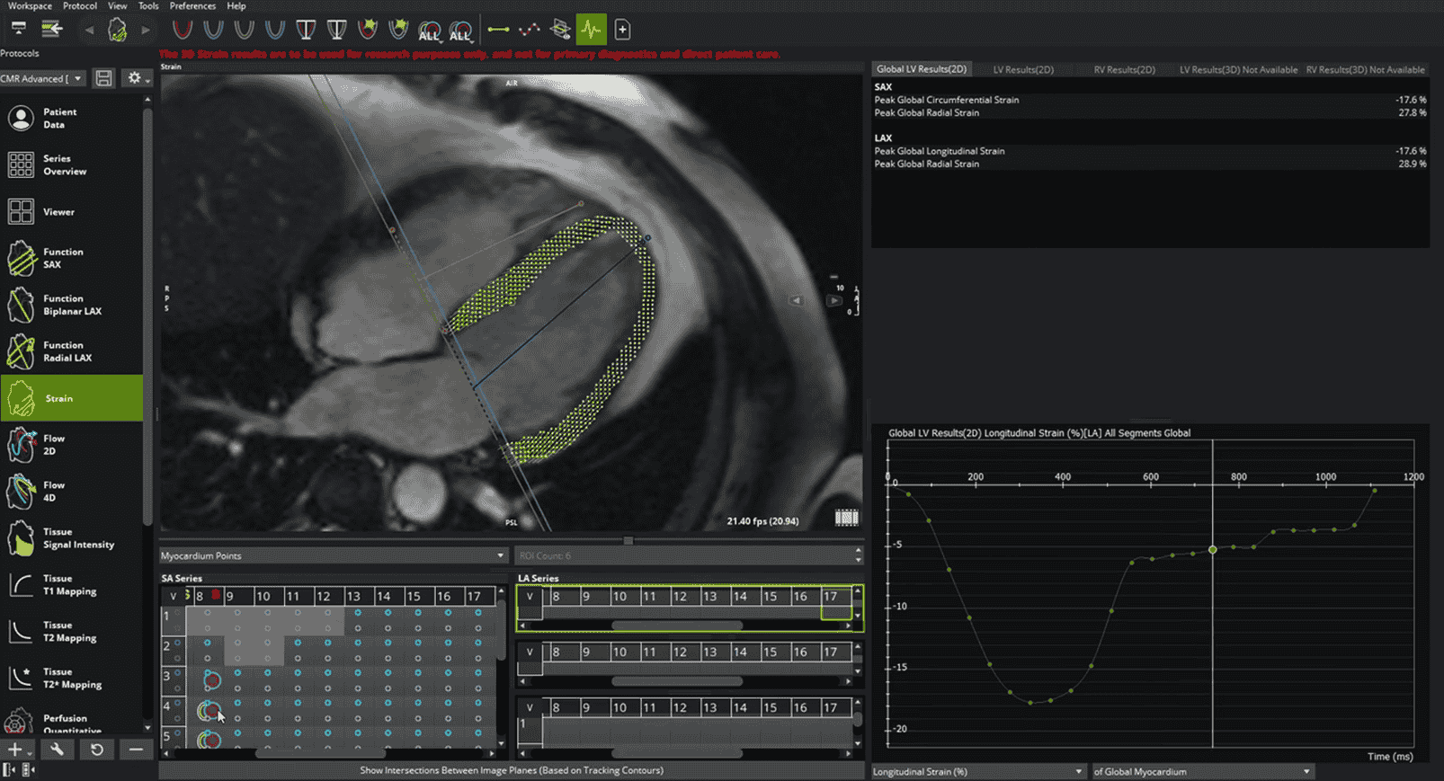
cvi42 provides strain analysis of MR images by providing measurements of 2D LV myocardial function along with measurements of 2D RV and 3D myocardial function. The software allows for tissue characterization of cardiac MR Images and perfusion analysis of cardiac Images and includes flow quantifications based on velocity encoded cardiac 2D images. It supports clinical diagnostics by quantitative measurement of the heart and adjacent vessels and by using area and volume measurements for measuring LV function and derived parameters.4
4D Flow
cvi42 allows simultaneous acquisition of 3D morphology and time-resolved blood flow velocities in three spatial directions. This module enables a variety of options for visualizations of complex blood flow patterns. It also facilitates the retrospective quantification of flow parameters with its flexible plane placement function.4
- DICOM-compliant SAX and/or LAX cine SSFP CRM image.
- As per scanning guidelines from the Society for Cardiovascular Magnetic Resonance (SCMR).5
- Strain Module uses myocardial contours on cardiac MR short-axis (SAX) and/or long-axis (LAX) SSFP (or similar such as GRE) images to track the myocardium and derive myocardial deformation parameters.
- Function
- SAX: AI-based left and right ventricular contour detection
- SAX: Polar map display for LV wall thickening and motion
- Biplanar LAX: AI-based LV, LA and RA contour detection in all phases
- Biplanar LAX: Automatically calculated long axis strain values
- Radial Long Axis: LV assessment with an axial acquisition
- Radial Long Axis: Semi-automated contour detection
- Flow
- Flow 2D: AI-based aortic and pulmonary contour detection
- Flow 2D: Qp: Qs comparison
- Flow 2D: Apply offset and anti-aliasing correction
- Tissue
- Signal Intensity: Calculate scar and edema percentage with late enhancement and T2 weighted imaging
- Signal Intensity: AI-based contour detection
- Signal Intensity: Multiple algorithm options for quantifying scar pattern presentations
- T1 Mapping: Motion correction of T1 raw images
- T1 Mapping: Native and post-contrast T1 and ECV map generation and customizable color maps
- T1 Mapping: Automated loading and AI-based contour detection of native and post-contrast maps
- T2 Mapping: Global and regional analysis
- T2 Mapping: Motion correction of T2 raw images
- T2 Mapping: T2 map generation and customizable color maps
- T2 Mapping: Automated loading and AI-based contour detection of T2 map
- T2 Mapping: Global and regional analysis
- T2* Mapping: Global and regional T2* analysis
- T2* Mapping: T2* color overlay
- T2* Mapping: Report of Iron content
- Semi-quantitative Perfusion: Qualitative viewer for rest and stress imaging
- Semi-quantitative Perfusion: Polar map and curve display for signal intensity
- Radial Long Axis: LV assessment with an axial acquisition
- Radial Long Axis: Semi-automated contour detection
- Strain
- Quantify global and regional radial, circumferential and longitudinal strain in 2D
- AI-based LV contour detection
- Calculate strain rate, displacement, time to peak strain and displacement, velocity, torsion, and torsion rate
- 4D Flow
- Preprocessing including offset correction and antialiasing
- Centerline definition for multiple structures
- Various flow visualizations
- Flow assessment in multiple planes Qp: Qs comparison
- Quantitative perfusion
- Streamlined workflow for rest and stress perfusion quantification
- Automated motion correction and contour detection
- Color map display of myocardial blood flow (MBF) values
- Color map display of myocardial perfusion reserve (MPR) values
- Vendor neutral with multi-sequence support
cvi42 offers customizable structured reporting for cardiac MR evaluation. Build the report you need for daily clinical practice.
Standard Integrated Report
- Simple Clinical data reporting
- Customizable template builder
- Infographics, diagrams, screenshots and measurements
- Export PDF, text, DICOM secondary capture and DICOM encapsulated pdf
- User configurable normal values and automatic classification
- Updated normal values sourced from the Healthy Hearts Consortium6
- Configure consistent findings text including values and results
- User permissions configuration
cvi42 Report
- Includes all standard reporting functionalities
- Z-Scores
- Advanced Database Search
- Browser Access
- HL7 Integration*
* Additional license required. HL7 integration requires the purchase of HL7 Integration Services.

Vendor
Circle Cardiovascular Imaging
EU risk class and CE marking
cvi42 has CE marking (CE2797) and risk class IIa
Reimbursement status
Not reimbursed
Target Population
The target population for cvi42 is not restricted, however cvi42’s semi-automated machine learning algorithms are intended for an adult population.
Contradictions
No contradictions given
Limitations
- The Strain Module implements an algorithm for modeling deformation from a two-dimensional (2D) version of the nearly incompressible deformable model and provides reasonably accurate estimates of the global strain measures. However, similar to other devices with such application it poses the following limitations:
- The accuracy of the estimates is limited by the image acquisition, image quality, pixel resolution, temporal resolution, presence of artifacts and noise.
- Results are dependent on the contours; the accuracy can be improved by providing contours in more phases.
- There is no established gold standard currently available across vendors and modalities; variability in results may exist comparing with different vendors or modalities.
- Plaque assessment results may differ between whole vessel or stenosis area selection. It is recommended to select smaller region that avoids intersections or bifurcations.
- Plaque may be incorrectly identified in the center of the vessel. Sculp tool can be used to restore the lumen in the region.
- Removing control points from the centerline can lead to varying results.
Other Offerings in the Cardiac Service Line
Discover Circle cvi42
on Calantic


